Dr. Jenny Gandhi

procedure steps

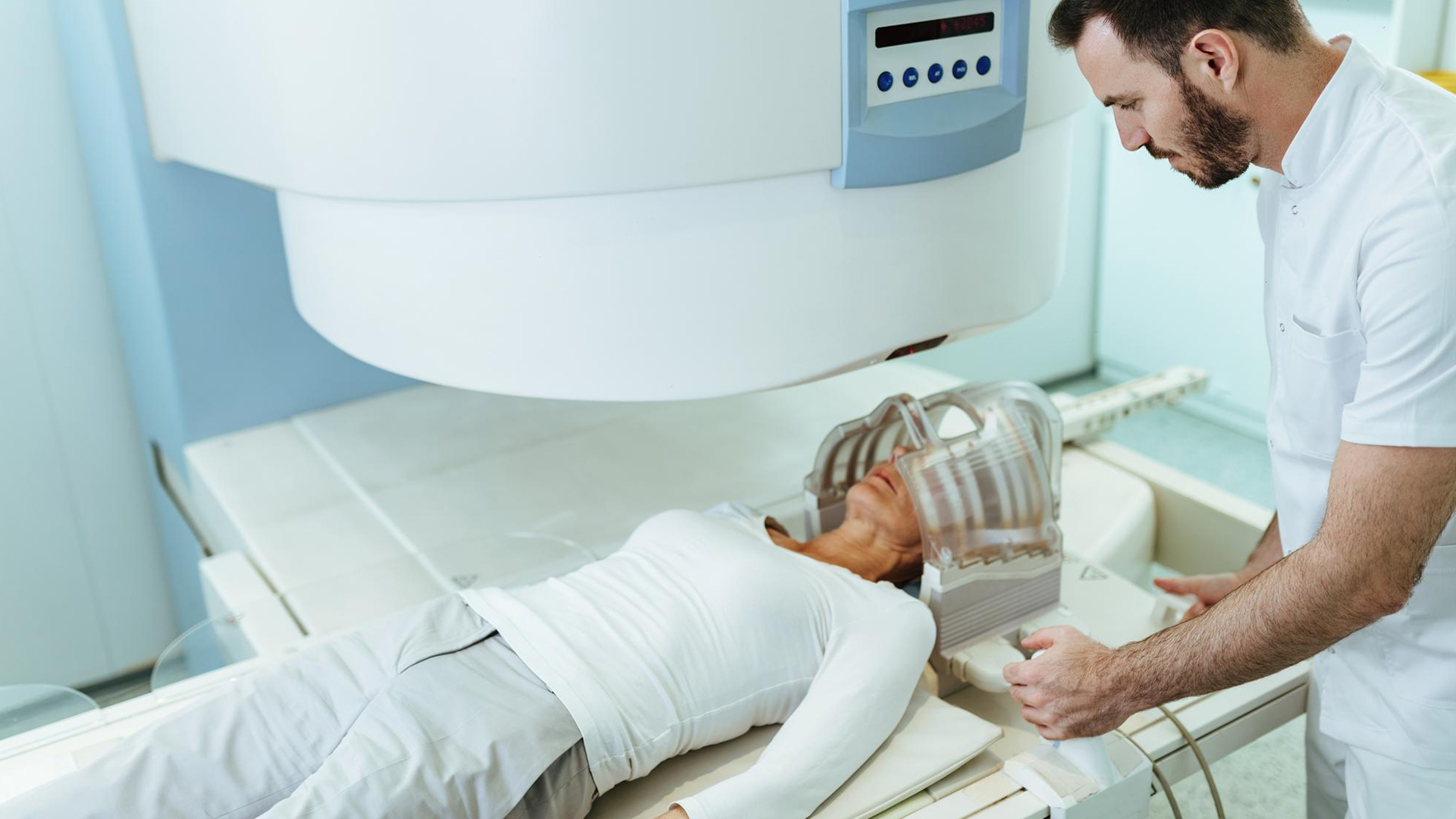
1. Positioning and Immobilization

To precisely immobilize and pinpoint the target area, local anesthetic is used to connect a stereotactic frame to the patient's head. In certain situations, immobilization may be accomplished with a bespoke thermoplastic mask rather than a frame.

2. Target Localization and Imaging

While the frame or mask is in place, high-resolution imaging, such as MRI, CT, or angiography, is carried out to locate and identify the target lesion. To precisely define the treatment area in three dimensions, image fusion integrates various scan modalities.

3. Planning for Treatment

The neurosurgeon and radiation oncologist create a radiation plan using specialized software. In order to accurately target the beams on the lesion while protecting the surrounding healthy tissue, this design calculates the radiation dose and angles.

4. Delivery of Radiation

The frame of the treatment couch is attached to the radiosurgery machine while the patient lies on it. In order to converge on the target, several narrow beams of concentrated radiation (like Gamma Knife or LINAC) are supplied from various angles. Throughout this often brief treatment, which can take anywhere from 30 minutes to several hours, the patient is kept awake and under observation.

5. Observation and Recuperation

Accuracy is ensured throughout treatment by motion monitoring and continual image guidance. The majority of patients return to their regular activities shortly after treatment, frequently the next day.

6. Follow-up

In the months that follow, routine follow-up imaging is carried out to evaluate the effectiveness of the treatment and schedule any further treatments that may be required.

disease treated

Brain Arteriovenous Malformation

Lorem Ipsum is simply dummy text of the printing and typesetting industry.

Spinal Vascular Malformation

Lorem Ipsum is simply dummy text of the printing and typesetting industry.

Dural Arteriovenous Fistulas

Lorem Ipsum is simply dummy text of the printing and typesetting industry.

suitable for

Functional Disorders and Trigeminal Neuralgia

Focused radiation treatment can be used to treat patients with trigeminal neuralgia or other neurological functioning disorders that are not improving with medication.

Difficult-to-Access or Inoperable Lesions

patients with lesions in important brain regions where surgery would be dangerous or inappropriate.

Good Life Expectancy and Performance Status

Those with good overall health, a reasonable life expectancy, and managed systemic disease such as brain metastases are ideal candidates.

Fewer Lesions

Instead of treating individuals with widespread disease, SRS works better for those with a small number of brain metastases (usually up to 3–4).

benefits

Treatment with Minimal Invasion

By delivering focused high-dose radiation without requiring surgical incisions, SRS lowers the risk of infection and hospital stays.

High Accuracy Targeting

SRS precisely targets cancers or lesions while minimizing side effects and protecting surrounding healthy tissue by using several converging radiation beams and sophisticated imaging.

Brief Treatment Time

usually finished in a single session or up to three to five sessions, enabling patients to avoid the drawn-out treatment regimens associated with traditional radiation.

Appropriateness for Inaccessible Lesions

useful for malignancies in eloquent or deep brain regions where surgery is impractical or dangerous.

Maintaining Neurological Function

SRS helps preserve neurological function and quality of life by reducing radiation exposure to healthy brain tissue.

Shorter Recuperation Period

Due to SRS’s low invasiveness and outpatient nature, patients frequently return to their regular activities soon after treatment.

High Rates of Tumor Control

For small to medium-sized tumors, AVMs, and functional problems, SRS provides excellent local control that is frequently on par with surgical results.

Reduced Risk of Complications

less adverse effects than open surgery or traditional whole-brain radiation, such as fatigue, nausea, or hair loss.

Adaptability in Handling Several Lesions

Convenience and efficacy can be increased by treating many tumors or targets in one session.

post-treatment recovery

Timeline for Recovery
After therapy, the majority of patients return to their regular activities within a few days. Although they usually go away in a few days to weeks, mild side effects including exhaustion, nausea, or scalp soreness close to frame pin locations can happen.
Handling Adverse Reactions
It's crucial to take breaks when necessary while continuing to engage in mild exercise, such as walking, as fatigue may persist for several weeks.
Over-the-counter painkillers can be used to treat pin site tenderness or swelling, which typically goes away in one to two days.
Discharge and Monitoring
Patients are frequently released the same day or following a short observation period.
About a month after treatment, follow-up appointments are scheduled to evaluate symptoms and may involve imaging to track the response of the tumor or lesion.
Long-Term Results
It can take months to years for a tumor to diminish; benign tumors usually take 18 months to two years, malignant tumors take a few months, and AVMs take longer.
Frequent MRI/CT scans track development, and if more treatments are required, they can be scheduled.
Assistance and Observation
Optimal healing and management of any complications are ensured by close multidisciplinary follow-up, which includes meetings with radiation oncologists and neurosurgeons.

Schedule Your
Consultation Today!