Dr. Jenny Gandhi

procedure steps

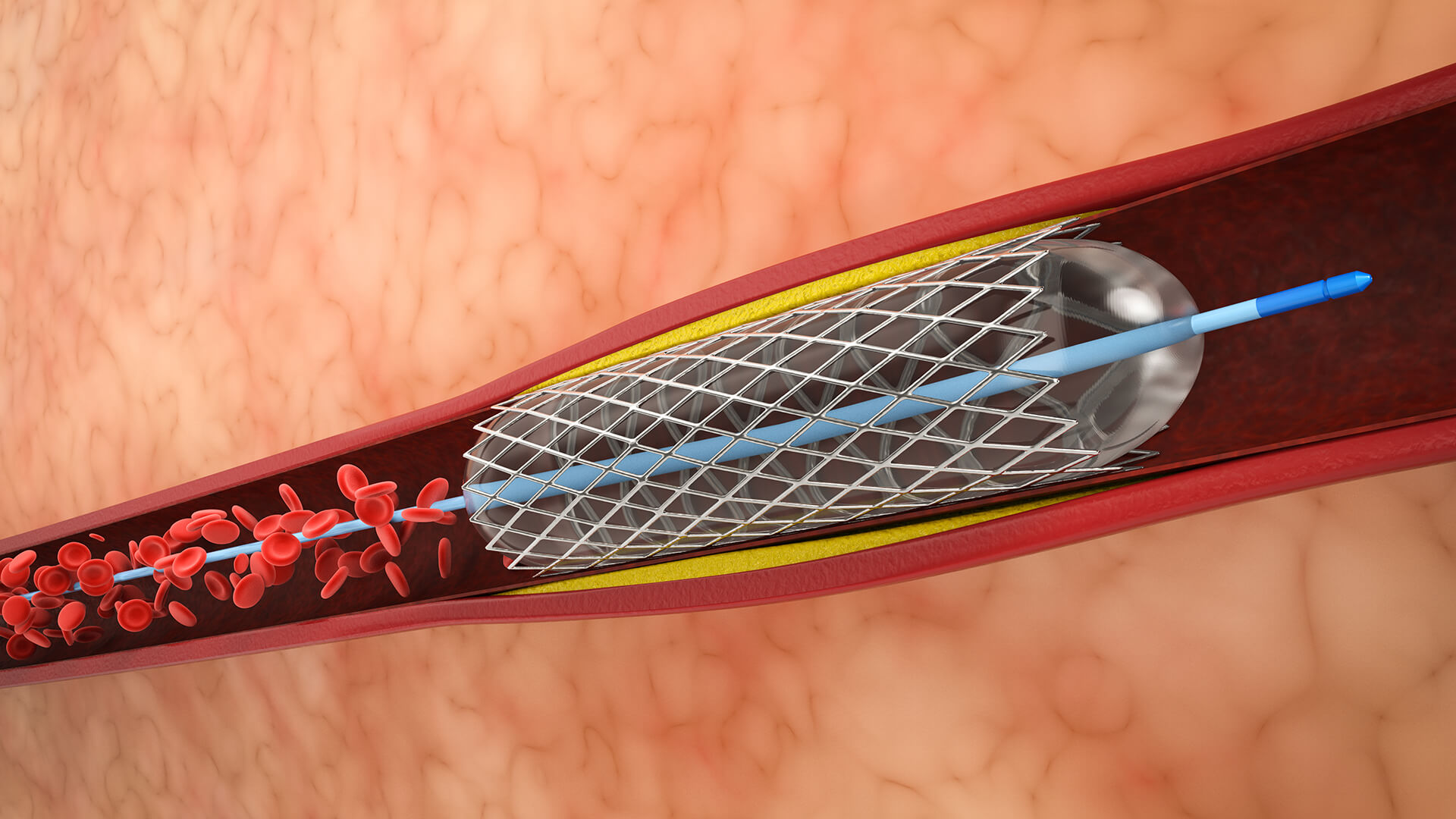
1. Vascular Access and Configuration

Obtain vascular access, usually through the femoral artery. In the parent artery that supplies the aneurysm, place a guiding catheter proximally.

2. Navigation of Catheters

Handle two microcatheters: one that has a compliant balloon placed across the neck of the aneurysm, and another that has been inserted into the aneurysm sac to supply coils.

3. Initial Coil Deployment and Balloon Positioning

Place the balloon microcatheter exactly across the neck of the aneurysm. With the balloon deflated, insert the first framing coil into the aneurysm sac.

4. Inflation of the Balloon During Coil Deployment

To avoid coil herniation into the parent artery and to change the form of the neck to enable safe coil packing, momentarily inflate the balloon across the aneurysm neck.

5. Coil Stability and Balloon Deflation Verify

Before extracting coils, regularly deflate the balloon to ensure coil stability and sufficient aneurysm occlusion.

6. Last Angiography

Perform final angiograms to verify total aneurysm occlusion and parent and branch vessel patency once coil packing is good.

7. Hemostasis and Catheter Removal

After verifying that the coiling device has stopped working, carefully remove the catheters and make sure the vascular access site is hemostatic.

8. Handling of Issues

In the event of a vascular rupture, be ready to reinflate the balloon right away, handle thromboembolic events quickly, and make sure the procedure is monitored.

disease treated

Spinal Vascular Malformation

Lorem Ipsum is simply dummy text of the printing and typesetting industry.

Brain Aneurysm

Lorem Ipsum is simply dummy text of the printing and typesetting industry.

suitable for

Individuals with small to medium-sized cerebral aneurysms

especially those with a neck diameter of less than 5 mm and a dome-to-neck ratio of at least 2:1.

Both ruptured and unruptured aneurysms

where coil implantation is favored by anatomy and endovascular access is possible.

Patients with increased surgical risk due to age

comorbidities, or aneurysm location in difficult-to-access brain areas.

Anterior and posterior circulation aneurysms

particularly those that can be treated endovascularly.

Those for whom open surgery is contraindicated

for whom surgical clipping is deemed more risky.

Patients with aneurysms

Patients with aneurysms should prioritize avoiding invasiveness and maintaining brain function.

Situations where emergency care is required

Situations where emergency care is required, such as aneurysmal subarachnoid hemorrhage.

benefits

Less intrusive than open surgical clipping, resulting in a speedier recovery and lower morbidity.

Efficient in stopping cerebral aneurysms from rupturing or bleeding again.

Appropriate for patients with aneurysms in surgically challenging areas or those at high surgical risk.

Both ruptured and unruptured aneurysms can undergo this procedure.

Permits the parent vessel and nearby branch vessels to be preserved.

Shorter operation duration and lower need for anesthesia.

Repeatable in the event of an aneurysm recurrence.

Reduced overall rates of complications when compared to surgical cutting.

In particular, balloon-assisted coiling reduces the risk of coil prolapse and maintains artery integrity while improving coil stability, packing density, and safety in wide-neck aneurysms.

Compared to stent-assisted methods, it typically reduces the need for long-term antiplatelet medication.

post-treatment recovery

Monitoring at the Hospital
In order to monitor the neurological and vascular access sites closely and identify any issues early, patients usually remain in the hospital for one to two days following the treatment.
Early Recuperation
If stable, the majority of patients can return home in one to three days.
Fatigue, mild headaches, and little discomfort at the access site are frequent and usually go away fast.
Limitations on Activities
Over the next few weeks, a gradual return to regular activities is recommended.
To promote vascular healing, hard lifting and strenuous activity should be avoided for a few weeks.
Imaging Follow-Up
In order to establish aneurysm occlusion and check for coil compaction or recurrence, imaging follow-up with angiography or MRI is typically conducted within one to six weeks.
Extended Recuperation
It takes one to three weeks to fully recover to a normal living range, depending on personal circumstances and complexity.
For months or years, ongoing follow-ups are conducted to track the status of aneurysms.
Possible Rehabilitation
Rehabilitation requirements are rare, however they would be necessary if there were neurological impairments or treatment-related issues.
Lifestyle and Medication
Patients should follow lifestyle change advice, such as controlling blood pressure and quitting smoking, and may require antiplatelet medication if adjunct devices, such as stents, were utilized.

Schedule Your
Consultation Today!