Dr. Jenny Gandhi

procedure steps

1. Preparing for the Procedure

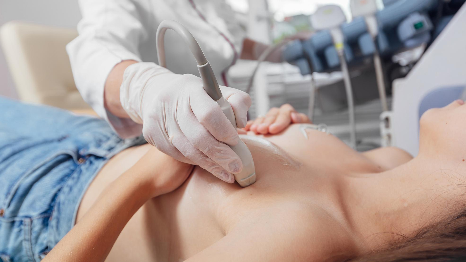
Use ultrasound imaging to confirm the location, size, and features of the abscess. Get the patient's informed permission after outlining the procedure, its dangers, and its advantages.

2. Sterilization and Positioning of Patients

Place the patient in a comfortable position so that the abscess site is easily accessible Use an antiseptic solution to clean and sanitize the skin surrounding the abscess.

3. Anesthesia locally

To numb the area surrounding the intended puncture site, apply local anesthetic (such as 1-2% lidocaine).

4. Localization of Ultrasound

To determine the best puncture location and provide real-time guidance for needle placement, use an ultrasound probe.

5. Needle Insertion and Aspiration

Using ultrasound guidance, insert a needle of the appropriate gauge (usually 16–18 gauge) into the abscess cavity. Aspirate all of the pus using a syringe; the fluid is sent for sensitivity testing and culture.

6. Repeat the aspiration If necessary

Determine whether any fluid is still visible on the ultrasonography. Until the cavity is sufficiently drained, more aspirations may be made.

7. Compression and Dressing

To reduce bleeding and hematoma formation, apply little pressure once the needle is removed. Use a sterile dressing to cover.

8. Antibiotics and Post-Procedure Care

Prescribe the right antibiotics in accordance with the results of the culture and clinical judgment. Arrange for additional clinical and ultrasound assessments to track the clearance of the abscess.

disease treated

Breat Abnormalities

Lorem Ipsum is simply dummy text of the printing and typesetting industry.

suitable for

Patients with Ultrasound-Accessible Diagnosed Abscesses

Those with ultrasound-confirmed abscesses, such as those in the breast, abdomen, pelvis, or soft tissues where percutaneous access is possible.

Breast Abscesses: Lactational and Non-Lactational

Both types can be effectively managed with this less intrusive option, which has a high success rate compared to surgical incision and drainage.

Different Sized Abscesses

can treat abscesses of any size; effective draining has been documented for abscesses greater than three centimeters. Repeated aspirations or alternate therapies may occasionally be necessary for multiloculated abscesses.

Individuals Who Should Not Have General Anesthesia or Surgery

Particularly helpful for people who choose minimally invasive methods or for whom surgery is hazardous or impracticable.

Early-Stage Abscesses

Without Significant Tissue Damage or Skin Necrosis. Ideal for abscesses that don’t have extensive cellulitis or underlying skin necrosis; in these situations, traditional incision and drainage may be necessary.

Patients Needing Additional Drainage

Abscess resolution can be accomplished without open surgery by repeating the procedure several times with adequate patient tolerance.

Settings with Limited Resources

Because of its affordability and accessibility, it is preferred in environments with limited surgical facilities.

benefits

A Less Invasive Option to Surgery

minimizes stress and recuperation time by avoiding the need for an open surgical incision and drainage.

High Rate of Success

Cure rates of up to 91–100% have been reported in numerous investigations, indicating its effectiveness in draining abscesses, particularly those less than 5 cm.

Improved Cosmetic Results

minimizes scarring in comparison to surgical drainage, which is crucial in places that are sensitive to appearance, such as the breast.

Faster Recuperation and Reduced Healing Time

Compared to surgical methods, patients have shorter recovery times and faster elimination of infection and inflammation.

Preservation of Breastfeeding

Compared to surgery, there is less tissue damage and pain, allowing nursing mothers to continue nursing.

Controlling Aspiration in Real Time

By visualizing needle insertion and drainage sufficiency, ultrasound guidance lowers the likelihood of recurrence and incomplete drainage.

Repeatable and Suitable for Outpatients

In order to minimize hospital stays, the aspiration is typically done as an outpatient procedure that can be repeated if necessary.

Reduced Rates of Complications

Reduced risk of problems compared to conventional incision and drainage, including bleeding, infection, and fistula formation.

Economical

Less costly and requiring less hospital resources than surgical drainage.

post-treatment recovery

Improvement of Symptoms and Hospital Stay
Within two to ten days following the surgery, clinical improvement is typically observed.
Depending on the extent of the abscess and the patient's condition, hospital release occurs after five to nine days.
Time for Healing
The abscess cavity may take several weeks to completely or nearly completely resolve, usually between two and four weeks, but it may take longer depending on personal characteristics like alcohol consumption or diabetes.
Aftercare
To evaluate abscess clearance and identify recurrence, patients undertake clinical and ultrasound surveillance at regular intervals (e.g., 48 hours, 1 week, 2 weeks, 1 month).
If necessary, aspiration may be done again.
Management of Antibiotics
Infection management is ensured in conjunction with drainage by adjusting antibiotics depending on culture results.
Recurrence and Complications
Although minor problems like small collection or persistent abscess are common, they usually go away with additional aspiration or medicines.
Compared to surgical incision and drainage, recurrence rates are lower.
Resuming Regular Activities
Depending on the severity and individual healing, most patients recover with little discomfort and cosmetic problems, returning to their regular activities in a matter of days to weeks.

Schedule Your
Consultation Today!