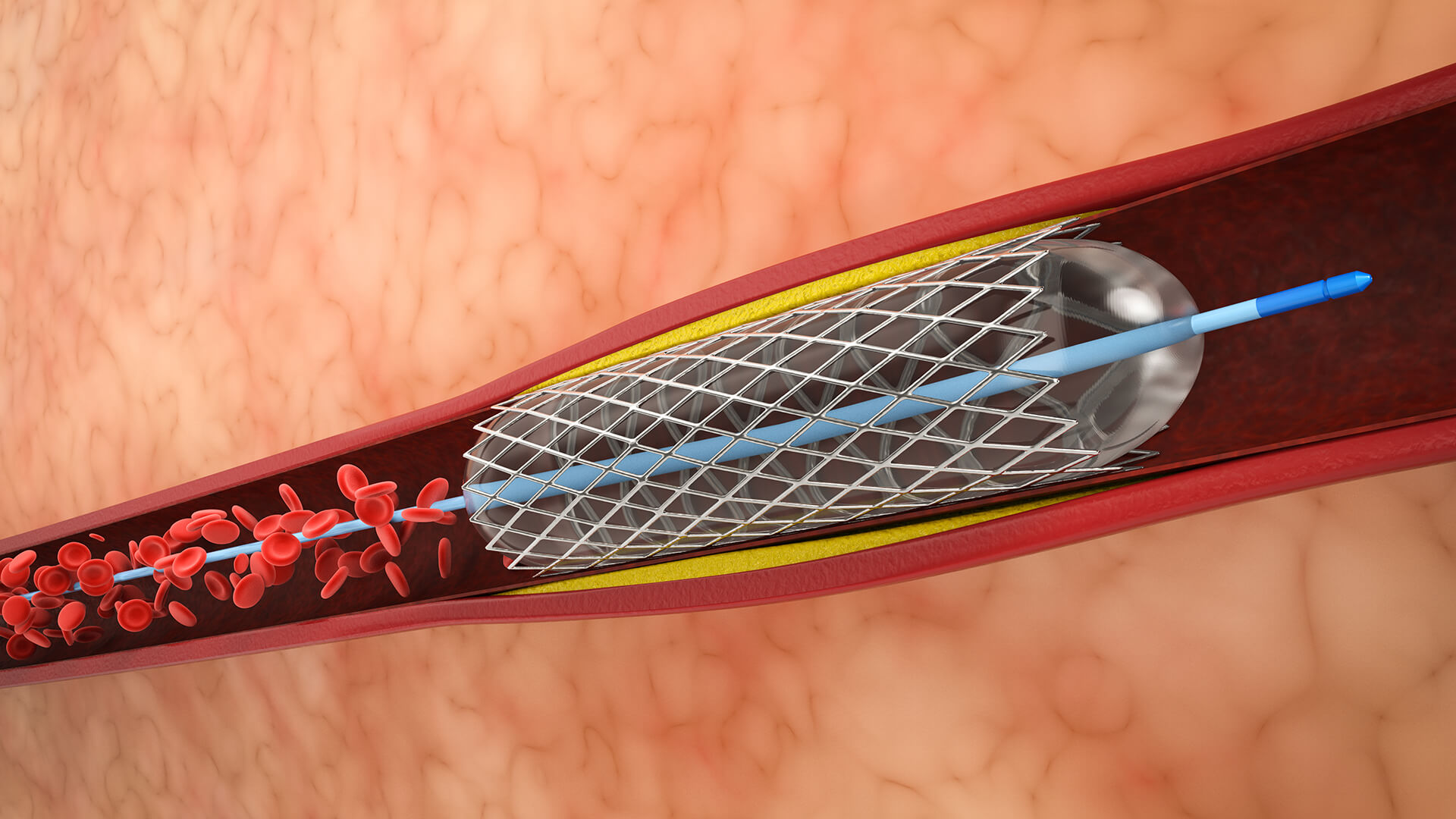
1. Preparation and Monitoring
In a sterile setting, the patient is ready. Sedatives and fluids are administered via an intravenous (IV) line. Heart rate, blood pressure, and oxygen level monitors are attached to the patient.

In a sterile setting, the patient is ready. Sedatives and fluids are administered via an intravenous (IV) line. Heart rate, blood pressure, and oxygen level monitors are attached to the patient.
The access site, which is typically in the arm, wrist, or groin, is made numb by applying local anesthetic. A sheath is inserted into the artery by a tiny puncture or incision.
Fluoroscopy is used to guide a tiny, flexible catheter with a deflated balloon at its tip through the sheath to the blocked or constricted artery.
To see the artery and pinpoint the precise location and degree of occlusion, contrast dye is administered via the catheter.
In order to expand the artery and restore blood flow, the balloon is inflated at the blockage site, compressing plaque against the arterial walls. This could cause considerable discomfort in the chest by momentarily reducing blood flow.
After the balloon is deflated and the catheter is removed, if necessary, a stent—a wire mesh tube—mounted on the balloon catheter is expanded in the artery to keep it open.
The sheath and balloon catheter are taken out. Either manually or with the use of a closure device, pressure is given to the access site to halt bleeding. A bandage is put on.
Vital signs and access site issues are tracked for the patient. While recovery times vary, most patients return to their regular activities in a matter of days.
Lorem Ipsum is simply dummy text of the printing and typesetting industry.
Ideal for people with severe coronary artery disease (CAD), which can cause symptoms including angina (chest discomfort), dyspnea, or decreased heart function because of constricted or blocked arteries.
In order to limit cardiac damage and rapidly restore blood flow, angioplasty is frequently carried out during or soon after a heart attack (STEMI).
Patients may benefit from improved blood flow and quality of life if their symptoms don’t go away despite changing their lifestyle and taking medicine.
When one or two arteries are blocked, angioplasty is recommended; bypass surgery may be taken into consideration for more severe disease.
those who are able to take antiplatelet drugs to avoid clot formation following stent implantation.
when there is a severe constriction that affects blood supply and causes symptoms.
Angioplasty is a less intrusive, quicker recovery option than open surgery.
By successfully opening constricted or blocked arteries and reestablishing blood flow to the heart or other afflicted organs, angioplasty reduces symptoms and guards against tissue damage.
It improves patients’ quality of life by reducing angina (chest pain), dyspnea, and exhaustion brought on by inadequate blood flow.
Timely angioplasty opens blocked arteries in acute situations, such as heart attacks, limiting damage to the heart muscle and reducing the chance of death.
Angioplasty is less invasive, requires fewer incisions, and typically results in shorter hospital stays and recovery times than coronary artery bypass surgery.
It is an effective therapeutic choice since blood flow is rapidly restored during the surgery, and most patients return to their regular activities in a matter of days.
Angioplasty improves overall cardiovascular health and allows patients to be more physically active by lowering symptoms
in conjunction with Stent Placement
By keeping arteries open for longer, stents lower the chance of additional constriction or blockages.


