1. Anesthesia and Preparation
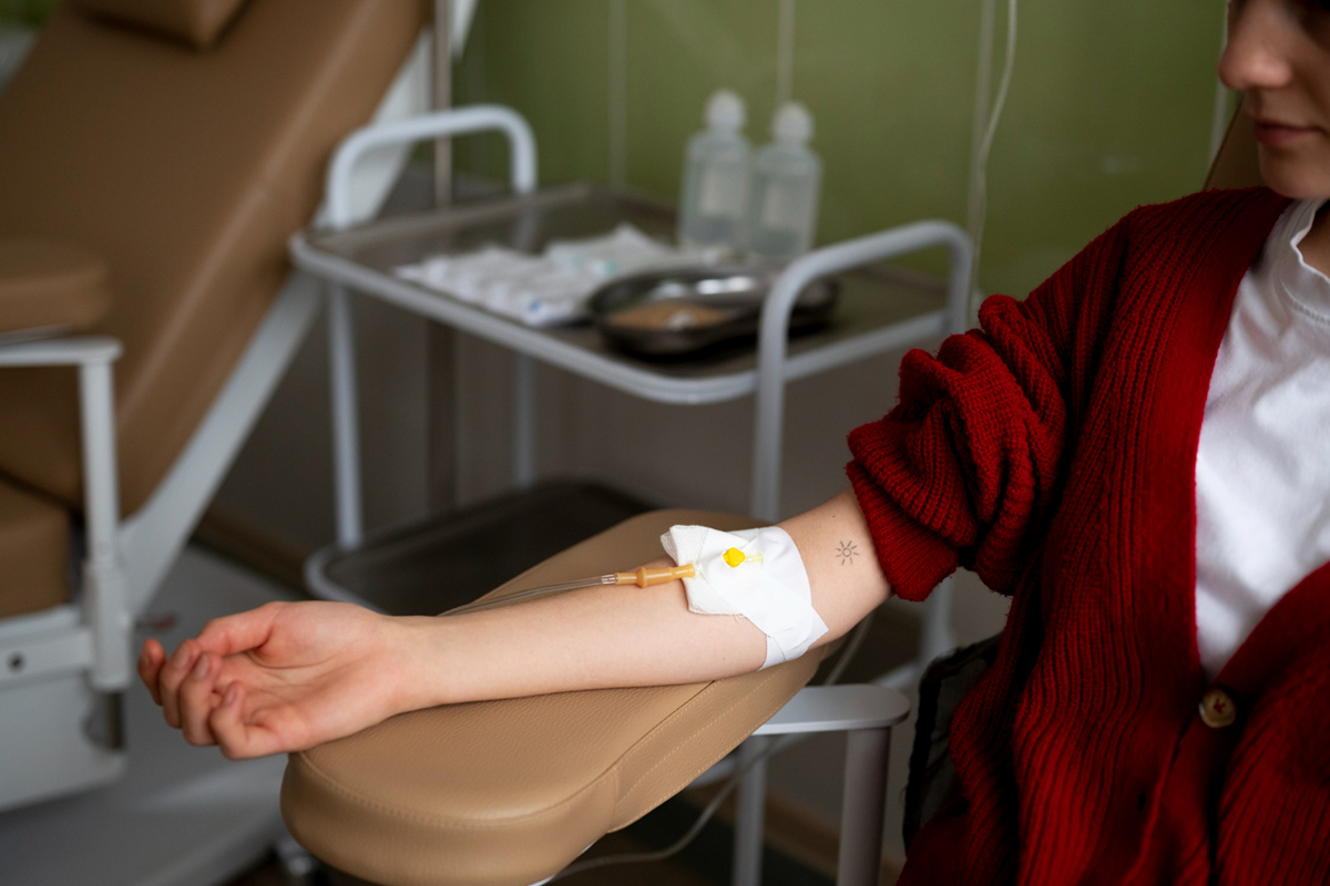
Local anesthetic is given at the vascular access location, which is typically the arm or groin, once the patient has been prepared. To help the patient relax, little sedation may be administered.


Lorem Ipsum is simply dummy text of the printing and typesetting industry.

Lorem Ipsum is simply dummy text of the printing and typesetting industry.