Dr. Jenny Gandhi

procedure steps

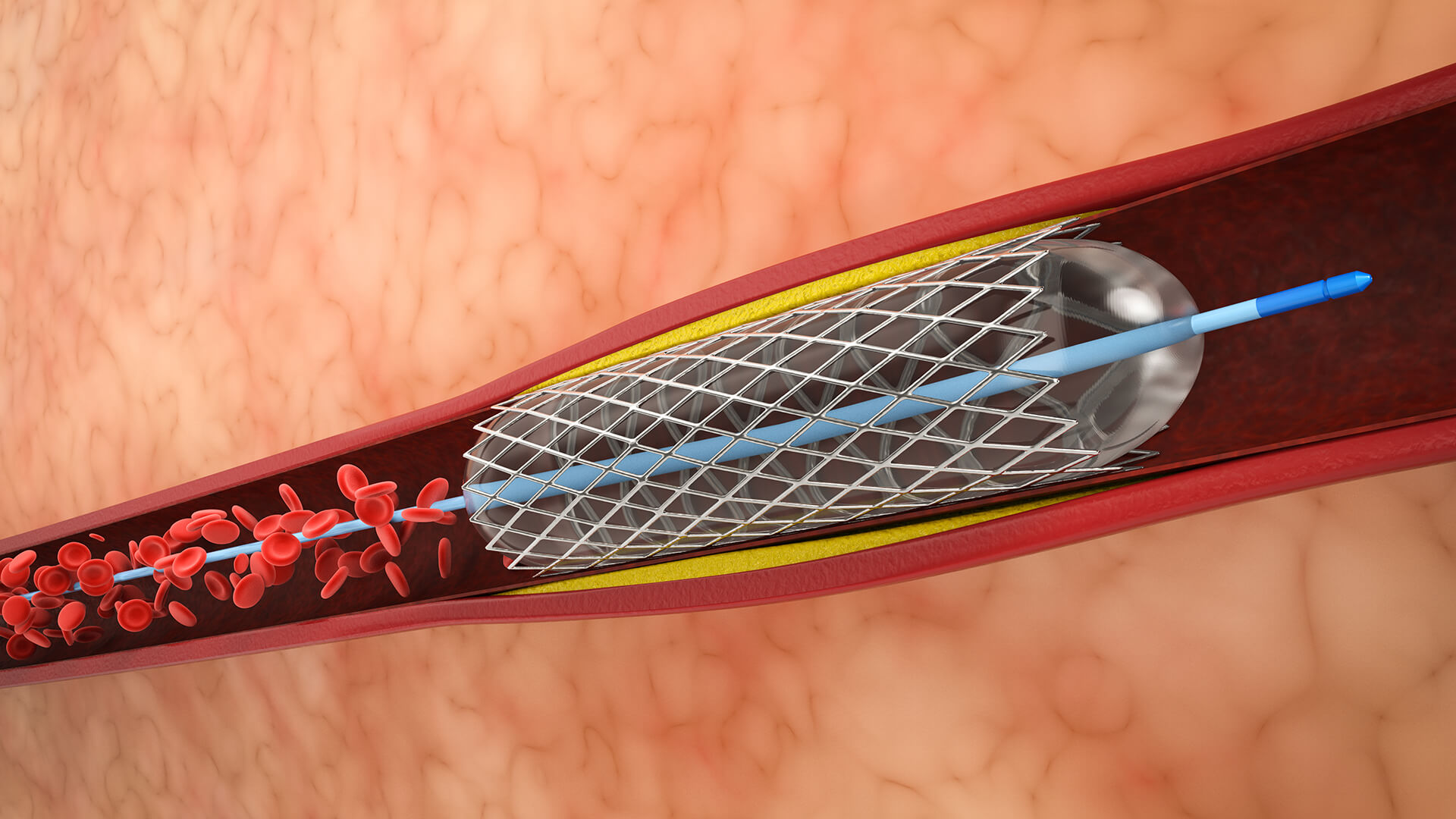
1. Placement of Catheters and Vascular Access

Usually, the femoral artery is used to get access. A guiding catheter is inserted close to the aneurysm in the parent artery.

2. Navigation of Balloon Microcatheters

The parent artery is used to navigate and place a compliant balloon microcatheter over the aneurysm's neck.

3. First Coil Installation

A different microcatheter is inserted into the sac of the aneurysm. To accomplish initial coil placement, the balloon is deflated before the first framing coil is deployed.

4. Inflation of Balloons (Remodeling)

In order to remodel the neck form and avoid coil herniation into the parent artery, the balloon is momentarily inflated across the aneurysm neck during coil deployment.

5. Balloon Deflation and Stability Assessment

To evaluate coil stability and make sure there is no prolapse prior to coil separation, the balloon is periodically deflated.

6. Last Angiography

Following final angiographic runs to verify total aneurysm closure and branch vessel preservation, balloons and microcatheters are removed after coil detachment.

7. Handling of Issues

In the event of a vascular rupture, tamponade is provided by immediate balloon reinflation. Pharmacological or mechanical therapies are used to quickly treat thromboembolic episodes or coil migration.

disease treated

Cerebral/venous Sinus Thrombosis

Lorem Ipsum is simply dummy text of the printing and typesetting industry.

Spinal Vascular Malformation

Lorem Ipsum is simply dummy text of the printing and typesetting industry.

Brain Aneurysm

Lorem Ipsum is simply dummy text of the printing and typesetting industry.

suitable for

Wide-necked cerebral aneurysms

particularly those that have a neck diameter of at least 4 mm or that include a sizable section of the parent artery.

Aneurysms

Aneurysms at risk of coil prolapse or herniation into the parent vessel due to anatomy.

ruptured and unruptured aneurysms

For both ruptured and unruptured aneurysms, maintaining the integrity of the parent vessel is essential.

unable to tolerate long-term dual antiplatelet medication

Patients who are unable to tolerate long-term dual antiplatelet medication are examples of situations in which stent-assisted coiling is less desirable.

anterior and posterior circulation aneurysms

Both anterior and posterior circulation aneurysms where it is difficult to secure coil implantation.

benefits

Prevents Coil Herniation

During deployment, the inflated balloon prevents coils from intruding into the parent artery by acting as a mechanical barrier across the aneurysm neck.

Neck Remodeling

Even in wide-neck or complex aneurysms, neck remodeling enables secure coil installation by temporarily changing and supporting the aneurysm neck.

Enhanced Coil Packing Density

Promotes more stable and dense coil packing inside the aneurysm sac, which may lower recurrence rates.

Versatility

Suitable for a broad range of aneurysm locations and morphologies, such as bifurcation and wide-necked aneurysms.

Decreased Need for Antiplatelet Therapy

Balloon-assisted coiling, in contrast to stent-assisted methods, typically requires less or no long-term dual antiplatelet therapy, which is advantageous for patients who are at risk of bleeding or have ruptured aneurysms.

Enhanced Safety

Enables tamponade by temporarily controlling blood flow in the event that an aneurysm bursts during the surgery.

Improved Visualization

Using angiographic contrast to better define the aneurysm neck during coil deployment increases procedural accuracy.

Instant Protection

During coil deployment, balloon inflation reduces procedural problems by acting as a safety mechanism.

post-treatment recovery

Hospitalization and Observation
After the procedure, patients typically remain in the hospital for one to two days in order to closely monitor their neurological condition and the location of their vascular access.
Early Recuperation Stage
It is normally possible to resume light activities in a few days. Patients may experience weariness, slight headaches, or discomfort at the access site, all of which are usually treatable with conventional pain medication.
Engagement and Attention
In order to promote vascular healing, patients are advised to refrain from physically demanding activities for a few weeks.
To stop bleeding or infection, the puncture site needs to be properly cared for.
Imaging Follow-Up
In order to verify the stability of the aneurysm occlusion and to look for any coil compaction or recurrence, follow-up angiographic imaging is usually planned one to six weeks after the treatment.
Long-Term Follow-Up
Regular clinical evaluations and imaging studies continue for up to months or years to monitor aneurysm status and to plan any additional treatment if necessary.
Outcomes and Prognosis
Most patients recover quickly compared to open surgery, with many returning to normal life and work within 1 to 2 weeks, depending on individual factors and complications.

Schedule Your
Consultation Today!