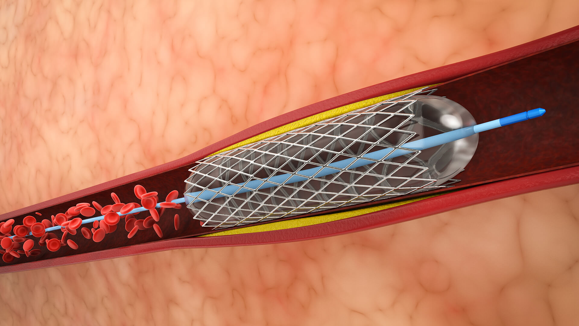
1. Access to Vascular
To reach a major artery, a little incision is usually made in the wrist or groin.

To reach a major artery, a little incision is usually made in the wrist or groin.
A catheter is inserted into the blood vessels up to the location of the brain artery blockage using real-time fluoroscopic (X-ray) guidance.
To place the device distal to the blockage, a microcatheter is inserted past the clot, frequently over a microwire.
An aspiration catheter or stent retriever is placed across or close to the clot. Aspiration catheters can suction the clot while the stent expands to engage and capture it.
Blood flow is restored by carefully pulling the stent retriever and trapped clot (or clot aspirated) back through the catheter.
If there is vascular narrowing or dissection, balloon angioplasty or stenting may be necessary.
To stop the bleeding, all catheters are taken out and the puncture site is compressed
The patient's neurological alterations, problems, and progress toward recovery are continuously monitored
Lorem Ipsum is simply dummy text of the printing and typesetting industry.
Individuals who experience an acute ischemic stroke due to a large vascular obstruction (LVO) in the internal carotid artery (ICA), basilar artery, or middle cerebral artery (MCA).
Ideally, candidates should come up within six hours of the onset of symptoms, while some patients may be eligible up to twenty-four hours later if advanced imaging reveals brain tissue that can be saved.
Individuals with a National Institutes of Health Stroke Scale (NIHSS) score of six or higher are considered to have moderate to severe neurological impairments
A substantial salvageable penumbra and mild early ischemia abnormalities, such as an Alberta Stroke Program Early CT Score (ASPECTS) of 6 or above, are examples of favorable imaging profiles.
Patients who were functionally independent before the beginning of the stroke (modified Rankin Scale score 0–2) were selected to maximize recovery potential.
For some people with basilar artery blockage, mechanical thrombectomy may also be helpful, depending on clinical and imaging assessments..
Blocked brain arteries can be swiftly opened by mechanical thrombectomy, resulting in instant reperfusion and reducing additional brain tissue damage.
greatly improves the likelihood of regaining independent function at 90 days, particularly when compared to thrombolytic treatment alone. One more patient will experience a successful neurological recovery for every four to six that are treated.
Even those who would not ordinarily be eligible for IV thrombolysis benefit from thrombectomy if they present up to 24 hours after the onset of a stroke and have favorable imaging revealing recoverable tissue.
Meta-analyses reveal a significant decline in the rates of death and disability, even among older adults, people with more severe strokes, and people who present to the hospital later.
For big vascular blockages (MCA, ICA, basilar), where systemic medications are less successful, thrombectomy is especially beneficial.
Thrombectomy increases the likelihood of minor impairment and recanalization rates when used as bridging therapy following IV thrombolytics.
Research shows comparatively low incidence of major adverse events, with the majority of hazards effectively controlled in centers with experience.


