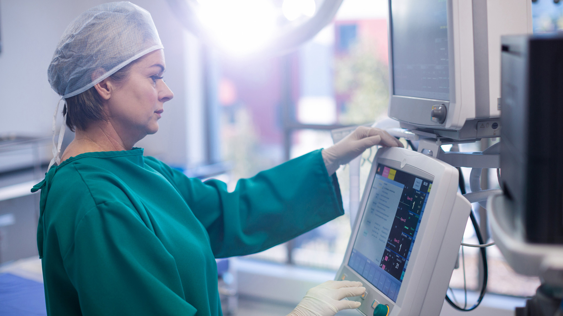
Dr. Jenny Gandhi

procedure steps

1. Getting Ready

Depending on the location of the tumor and the patient's condition, either general anesthesia or conscious sedation is given while the patient is positioned. To precisely detect the tumor, imaging guidance, typically an ultrasound or CT scan is put up.

2. Insertion of Needles

To ensure precise implantation, a narrow microwave antenna probe is gently inserted into the skin and moved into the tumor's center while being guided by real-time imaging.

3. Delivery of Microwave Energy

When the microwave generator is turned on, electromagnetic energy is produced, quickly heating the tumor tissue. Coagulative necrosis is brought on by temperatures above 60°C, which effectively kills tumor cells in a matter of minutes.

4. Observation and Modification

Imaging tracks the expanding necrotic zone surrounding the probe during ablation. To obtain full coverage, including a margin of surrounding healthy tissue to lower the chance of recurrence, energy supply and antenna position can be changed.

5. Finalization and Probe Elimination

The antenna is removed and the microwave energy is cut off once a sufficient ablation zone has been established. To reduce bleeding and tumor seeding, certain systems execute tract ablation during needle removal.

6. Imaging After Ablation

To confirm the degree of tumor elimination and look for problems, contrast-enhanced imaging (CT or ultrasonography) is usually done right away or soon after.

disease treated

Liver Cancer

Lorem Ipsum is simply dummy text of the printing and typesetting industry.

Renal Tumors

Lorem Ipsum is simply dummy text of the printing and typesetting industry.

Lung Tumors

Lorem Ipsum is simply dummy text of the printing and typesetting industry.

suitable for

Patients with Early-Stage Hepatocellular Carcinoma

According to Milan criteria, MWA is appropriate for patients with tiny liver tumors, usually a single tumor ≤5 cm or up to three nodules each <3 cm.

Individuals Unfit for Surgery

those who refuse surgery, have limited liver reserve, or have comorbidities that make them unsuitable for liver resection.

Location and Accessibility of the Tumor

tumors that are easily accessible and visible under CT or ultrasound guidance for accurate probe placement.

Sufficient Liver Function

For MWA treatment to be safe, patients typically need to have Child-Pugh class A or B liver function.

Excellent Performance Status

For patients to tolerate the surgery, their ECOG performance level should be between 0 and 1.

Exclusion Standards

Patients with portal vein thrombosis, extrahepatic metastases, significant tumor burden (>5-6 cm tumor), or severe liver decompensation are typically not candidates for MWA.

benefits

Procedure with Minimal Invasiveness

By doing MWA through a tiny skin puncture rather than open surgery, there is less stress, a lower chance of infection, and less pain following the procedure.

Tumor Destruction That Is Quicker and More Effective

Compared to previous ablation methods, MWA produces greater temperatures quickly (over 100°C), allowing for bigger and more reliable ablation zones in less time.

Capacity to Treat Multiple and Larger Tumors

Larger tumors (up to 5 cm or more) and several lesions at once can be safely treated with MWA, which is frequently difficult with other modalities.

Reduced Recuperation Time

With shorter hospital stays, less suffering, and a quicker return to regular activities, patients usually heal more quickly.

Decreased Blood Loss When Combined with Surgery

MWA can minimize intraoperative bleeding by closing blood arteries when administered in conjunction with liver resection.

Repeatable Therapy

MWA is a versatile method for managing cancer since it can be repeated if tumors relapse or new lesions appear.

Better Local Control and Survival

Research shows that MWA offers patients improved quality of life and strong tumor control with promising survival rates.

post-treatment recovery

Quick Recuperation
Following the treatment, patients typically stay in a recovery area for a few hours while their vital signs, level of pain, and overall health are monitored. Depending on their condition, some patients may be released from the hospital the same day, but the majority stay overnight for observation and pain control.
Symptoms Following Ablation
Localized pain, exhaustion, low-grade fever, and a flu-like syndrome with nausea and malaise are typical mild symptoms.
Although they can occasionally continue longer, these symptoms typically last one week. With supportive care and recommended pain medication, they are usually under control.
Limitations on Activities
For roughly seven days, patients are recommended to relax and refrain from physically demanding activities.
Within a week to ten days following the treatment, the majority of patients gradually return to their regular daily activities.
Monitoring Follow-Up
Four to six weeks following ablation, follow-up imaging is performed, usually using contrast-enhanced CT or MRI, to evaluate the extent of tumor eradication and track liver function.
Based on the imaging results, additional therapy or repeat ablation may be scheduled.
Complications
Serious problems, such as bleeding, infection, or harm to nearby organs, are rare.
Severe pain, fever, jaundice, or any other odd symptoms should be reported right away by patients.

Schedule Your
Consultation Today!