Dr. Jenny Gandhi

procedure steps

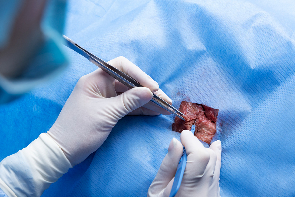
1. Setting Up and Positioning

Usually in a comfortable position, the patient lies down. Depending on how visible the lesion is, ultrasonography, mammography, or MRI guidance are used to pinpoint its location.

2. Anesthesia locally

To ensure a painless operation, local anesthetic is given around the biopsy site to numb the skin and tissue.

3. Making an incision and inserting a needle

A little (3–5 mm) skin incision is made. Through this incision, a specialized vacuum-assisted biopsy needle is introduced and precisely guided to the lesion being imaged.

4. Sampling of Tissue

Tissue is drawn into the needle chamber by the device's suction mechanism, and the tissue sample is removed by a revolving cutter. One needle insertion can be used to collect many samples.

5. Excision of Lesions

Excision biopsies minimize the necessity for surgical removal by sequentially sampling the whole lesion or aberrant tissue.

6. Placement of Optional Markers

If the lesion is entirely excised, a small titanium marker may be positioned at the biopsy site to pinpoint the area for future reference.

7. Dressing and Needle Removal

To stop any bleeding, the needle is removed and pressure is administered. The site of the incision is covered with a little bandage.

8. Recuperation and Observation

Patients usually return home the same day after a brief observation for bleeding or problems. Over-the-counter medications are used to manage the slight pain.

disease treated

Breat Abnormalities

Lorem Ipsum is simply dummy text of the printing and typesetting industry.

suitable for

Individuals with Benign Breast Lesions

Benign breast tumors such as fibroadenomas, tiny cysts, papillomas without atypia, radial scars, and other benign lesions that do not require surgical removal are best removed with VAEB.

Uncertain Malignant Potential Lesions

In order to minimize the necessity for open surgery, it is utilized for biopsies and the excision of high-risk or borderline lesions like papillomas, lobular neoplasia, flat epithelial atypia, and atypical ductal hyperplasia.

Individuals with Non-Palpable or Palpable Lesions

Suitable for lesions found by imaging that need to be diagnosed or removed, such as ultrasound-visible lesions and clustered microcalcifications under stereotactic guidance.

Individuals Requiring Bigger Tissue Samples

Compared to core needle biopsy, VAEB offers bigger, more representative tissue samples, improving diagnostic precision and lowering sampling error.

Patients Looking for Less Invasive Surgical Options

especially helpful for people who wish to avoid the risks, healing period, and scars that come with typical surgical excision.

Lesions that are small to medium

Lesions less than 2.5–3 cm are best suited for the device’s full excision.

Patients Needing a Second Biopsy or Tissue Diagnosis Prior to Treatment

This is helpful when previous biopsies were inconclusive or when clinical suspicion endures in spite of negative or unclear results.

benefits

Minimally invasive

VAEB causes less tissue damage and scarring because it only requires a tiny incision (3–5 mm) and does not require open surgery.

High Precision in Diagnosis

improves diagnostic accuracy, particularly for tiny, non-palpable, or microcalcified breast lesions, by enabling bigger, contiguous tissue samples with fewer needle insertions.

Possible Total Removal of the Lesion

Benign and some high-risk lesions can be fully removed with VAEB, frequently eliminating the need for further surgical excision.

Decreased Rates of Repeat Biopsies

Compared to traditional core needle biopsy, VAEB reduces the likelihood of false negatives and the need for repeat biopsies because of the quality and quantity of tissue collected.

Reduced Pain and Quicker Recuperation

Compared to surgical biopsy, patients recover more quickly and with less pain and suffering, enabling them to return to their regular activities sooner.

Improved Cosmetic Results

Compared to surgical excision, the precision method and little incision produce better cosmetic results with less scarring.

Advantages for the Mind

High patient satisfaction with less anxiety as a result of quicker diagnosis and treatment and less invasive techniques.

post-treatment recovery

Quick Recuperation
After the procedure, patients are usually released home the same day after a brief period of observation.
The biopsy site frequently experiences mild soreness or bruising, which normally goes away in two to three days.
Care of Incision
Adhesive strips or a little bandage are used to close the tiny incision, and it should be dry and clean for two to three days.
For the first twenty-four hours, patients are recommended not to take a bath or shower.
Bruising and Pain
Over-the-counter painkillers can be used to treat mild pain or a bruised feeling that may last for a few days.
Sometimes there is more severe bruising, which could take a few weeks to heal completely.
Limitations on Activities
To minimize bleeding and encourage healing, heavy lifting and strenuous exercise should be avoided for 48 hours following the surgery.
Resuming Regular Activities
Depending on their particular healing and the amount of the treatment, most patients return to their regular daily activities, including employment, within a few days to a week.
Results and Follow-Up
After tissue samples are sent for pathology, the results are usually ready in one to two weeks.
Follow-up appointments evaluate recovery and go over pathology findings.
Potential Issues and Surveillance
Hematomas and minor bleeding are uncommon problems that are typically treated conservatively.
If a patient experiences worsening pain, swelling, or other indications of an infection, they should seek medical attention.

Schedule Your
Consultation Today!