Brain Arteriovenous Malformation

Brain Arteriovenous Malformation
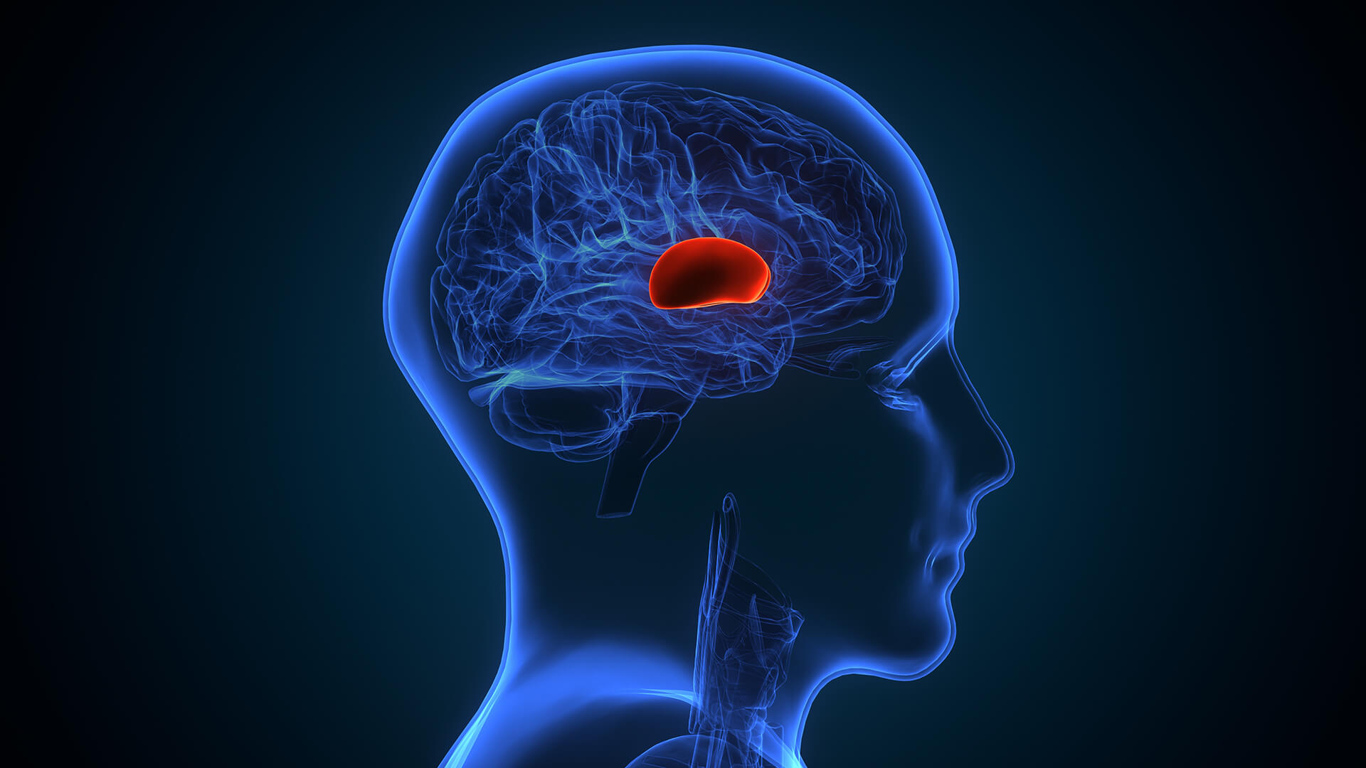
Brain hemorrhage can result in strokes, brain damage, or even death. Hemorrhage risk is between 2 and 4% annually, and it rises if the AVM has previously bled.
Decreased oxygen delivery to brain tissue, which can result in seizures, neurological impairments, and tissue damage.
Headaches, seizures, and neurological symptoms like weakness, numbness, or vision issues are brought on by pressure on nearby brain regions.
If left untreated, severe AVMs can be fatal or result in permanent disability.

diagnosis
Lorem Ipsum is simply dummy text of the printing and typesetting industry. Lorem Ipsum has been the industry's standard dummy text. Lorem Ipsum is simply dummy text of the printing and typesetting industry
Cerebral Angiography
-
 By carefully mapping the AVM's blood supply using contrast dye injection, cerebral angiography provides fine-grained images of blood vessels.
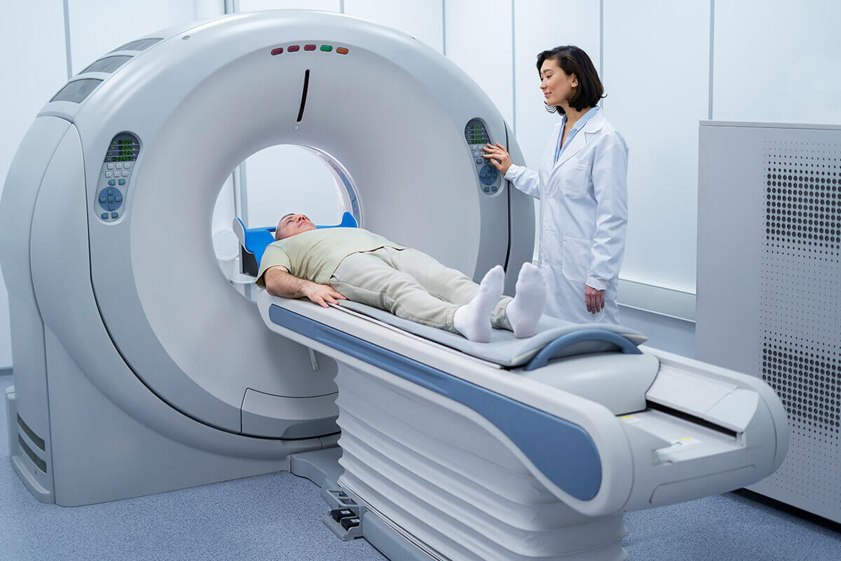
CT/CT Angiography
-
Shows abnormal vessel formations, bleeding, and brain structure using X-rays and contrast.
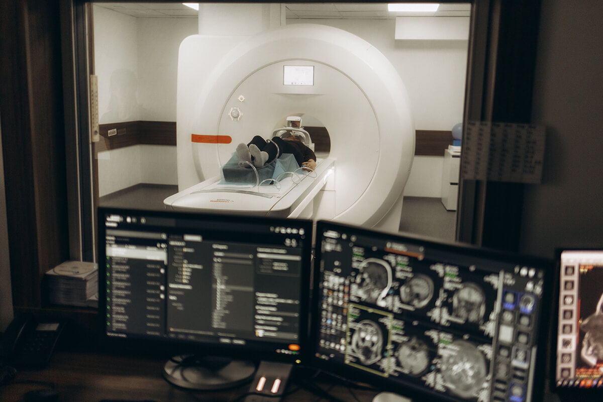
MRI/MR Angiography
-
To locate and define the AVM, MRI/MR Angiography provides fine-grained images of the brain's tissue and blood flow patterns.
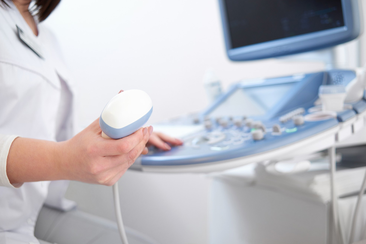
Transcranial Doppler Ultrasound
-
Identifies aberrant high-velocity patterns linked to AVMs by measuring blood flow in the brain arteries.