Adenomyosis

Adenomyosis


Progressive worsening of symptoms such as heavy menstrual bleeding and severe pelvic pain.
Chronic iron-deficiency anemia due to persistent heavy bleeding, causing fatigue, weakness, and shortness of breath.
Infertility or difficulty conceiving, as the abnormal uterine lining may interfere with embryo implantation.
Increased risk of miscarriage and complications in pregnancy such as preterm birth and small for gestational age babies.
Enlargement of the uterus, sometimes causing a protruding abdomen ("adenomyosis belly") and pelvic pressure symptoms.
Reduced quality of life due to chronic pain and emotional distress, including potential anxiety or depression.
In severe cases, hysterectomy (surgical removal of the uterus) may become necessary for symptom relief.

diagnosis
Lorem Ipsum is simply dummy text of the printing and typesetting industry. Lorem Ipsum has been the industry's standard dummy text. Lorem Ipsum is simply dummy text of the printing and typesetting industry
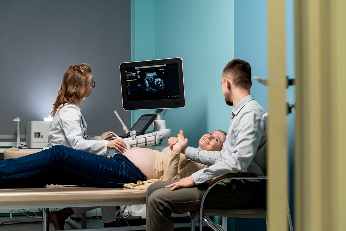
Transvaginal Ultrasonography (TVUS)
-
 The first-line imaging technique is transvaginal ultrasonography (TVUS), which is accessible and reasonably priced. A globular uterus, asymmetrical myometrial thickening, fan-shaped shadowing, and an irregular or interrupted junctional zone are some of the characteristics that may be present. Its specificity may be constrained, though.
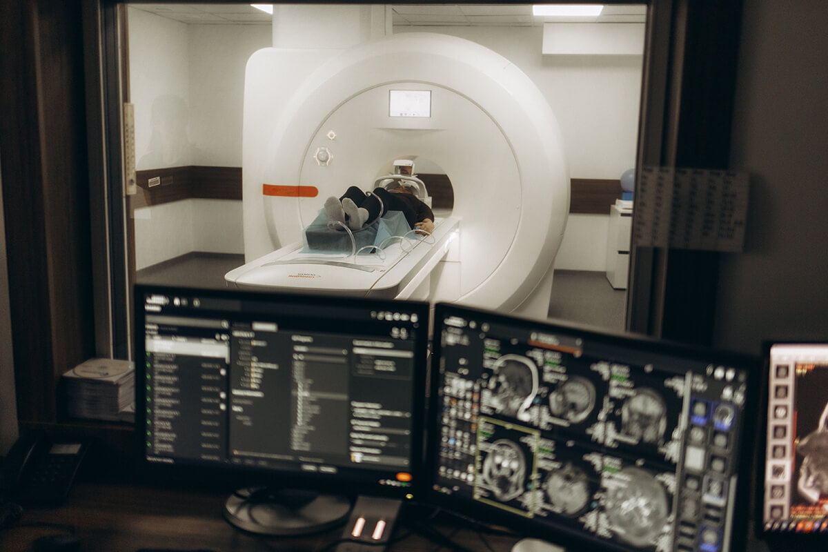
Magnetic Resonance Imaging (MRI)
-
When diagnosing adenomyosis, magnetic resonance imaging (MRI) has a higher sensitivity and specificity than TVUS. High-intensity myometrial foci, junctional zone thickening (>12 mm), and the ability to differentiate between focal and diffuse adenomyosis can all be clearly seen with MRI. When ultrasound results are unclear or complex, it is particularly helpful.
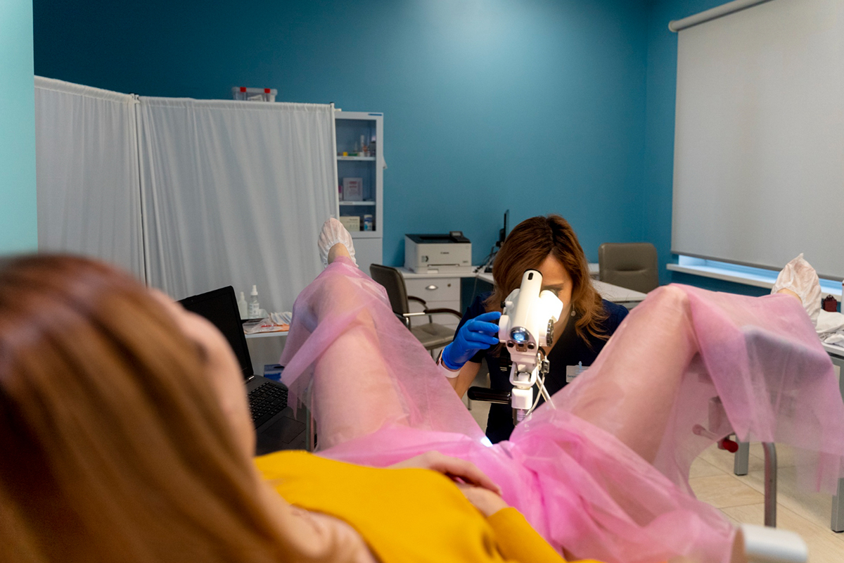
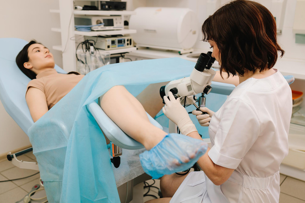
Hysteroscopy
Histopathology
-
Although histological examination is rarely done purely for diagnosis prior to treatment, it is confirmed to show ectopic endometrial tissue within the myometrium following hysterectomy.