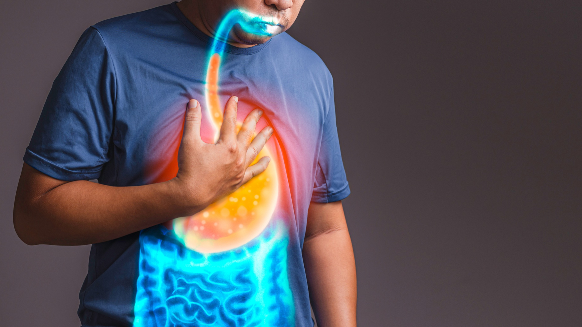
Ascites Or Pleural Effusion

Ascites Or Pleural Effusion

Spontaneous bacterial peritonitis (SBP): Infection of the ascitic fluid causing fever, abdominal pain, and sepsis, which can be life-threatening without prompt treatment.
Hepatic hydrothorax: Fluid buildup in the pleural cavity due to ascites, leading to breathlessness and chest discomfort.
Electrolyte imbalances and kidney failure, including hepatorenal syndrome, a type of acute kidney failure in cirrhosis patients.
Ascites-related hernias due to increased abdominal pressure.
Malnutrition and weight loss from early satiety and impaired protein synthesis.
Gastrointestinal bleeding and bowel complications in advanced cases.
Poor prognosis with high mortality rates; roughly 50% of patients with ascites from cirrhosis may die within 5 years.
Decline in quality of life due to abdominal discomfort, difficulty breathing, and fatigue.

diagnosis
Lorem Ipsum is simply dummy text of the printing and typesetting industry. Lorem Ipsum has been the industry's standard dummy text. Lorem Ipsum is simply dummy text of the printing and typesetting industry

Physical Examination
-
 Checking for abdominal distension, shifting dullness, and fluid waves, though noticeable only when fluid volume is significant.
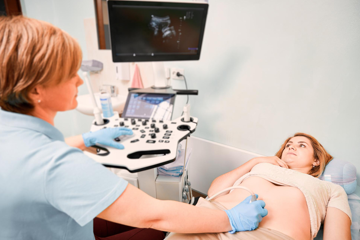
Ultrasound Imaging
-
The most sensitive and widely used technique to detect even small amounts of ascitic fluid, assess liver and abdominal organ status, and guide fluid drainage.
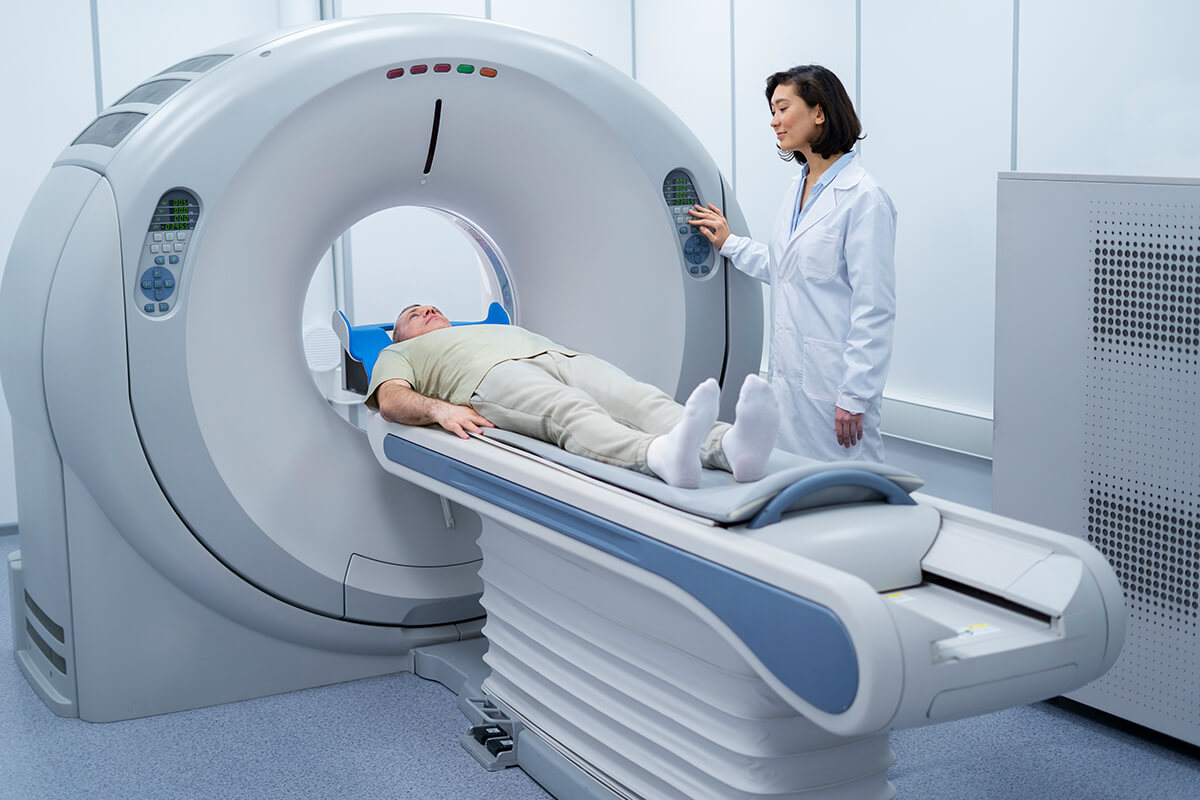
CT Scan
-
Provides detailed images of abdominal structures, useful for assessing organ abnormalities or masses causing ascites.

Diagnostic Paracentesis
-
Insertion of a needle to aspirate ascitic fluid for analysis. This is the gold standard for confirming ascites cause and ruling out infection like spontaneous bacterial peritonitis (SBP).
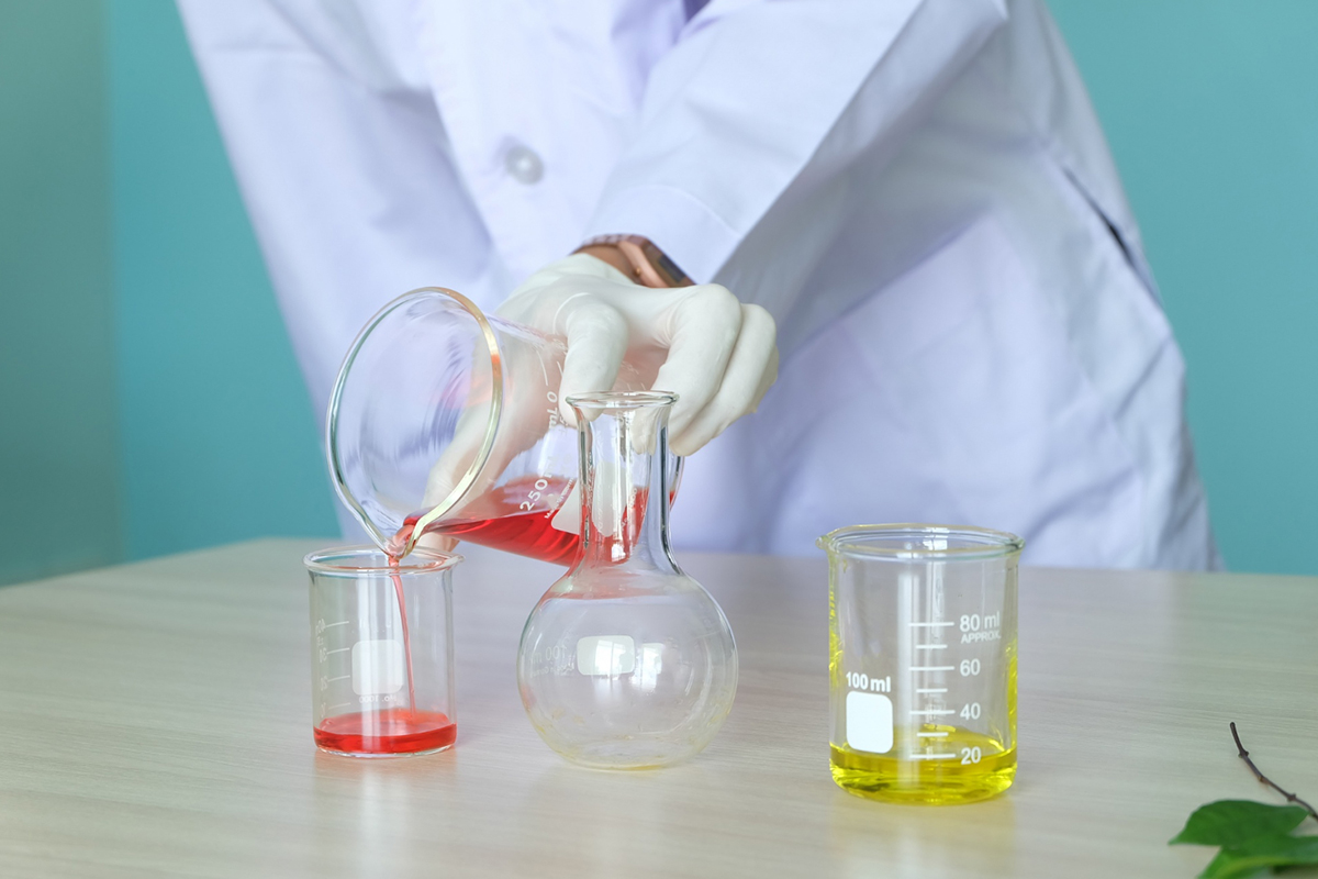
Laboratory Analysis of Ascitic Fluid
-
Includes cell count, protein level, albumin concentration to calculate serum-ascites albumin gradient (SAAG), culture for infection, cytology for malignancy, amylase for pancreatic origin, and other specific tests based on clinical suspicion.

Blood Tests
-
Liver function tests, kidney function, and coagulation panels to assess underlying conditions causing ascites