Deep Vein Thrombosis

Deep Vein Thrombosis


A life-threatening condition where a clot breaks free and blocks blood flow to the lungs.
Chronic pain, swelling, skin discoloration, and ulcers caused by vein damage from the clot.
Poor blood flow through veins leading to persistent leg swelling and skin changes.
Increased risk of developing another blood clot.
Rare but severe tissue death due to blocked blood flow, potentially leading to amputation.

Diagnosis
Lorem Ipsum is simply dummy text of the printing and typesetting industry. Lorem Ipsum has been the industry's standard dummy text. Lorem Ipsum is simply dummy text of the printing and typesetting industry

Physical Examination
-
 The healthcare provider checks for swelling, tenderness, and skin color changes in the leg.
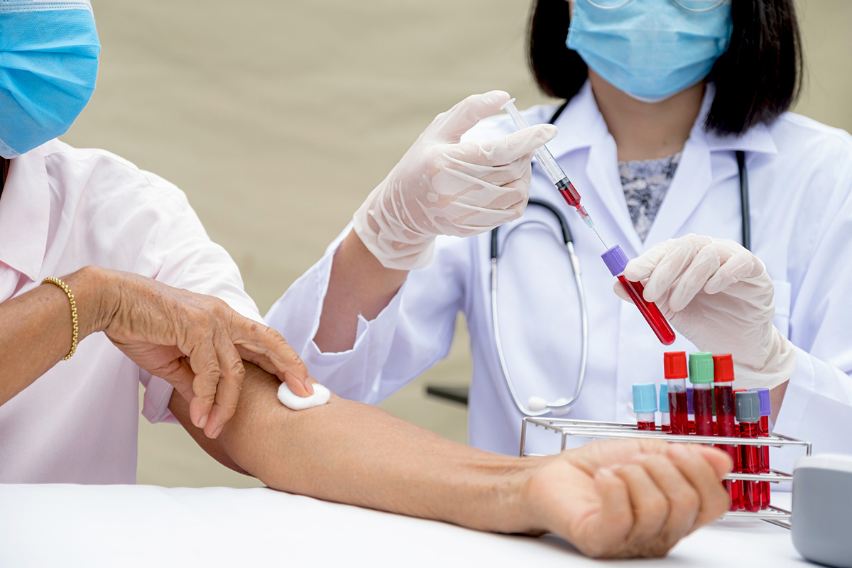
D-dimer Blood Test
-
Measures a substance released when blood clots dissolve. A high level suggests the possibility of a clot but is not specific to DVT alone.

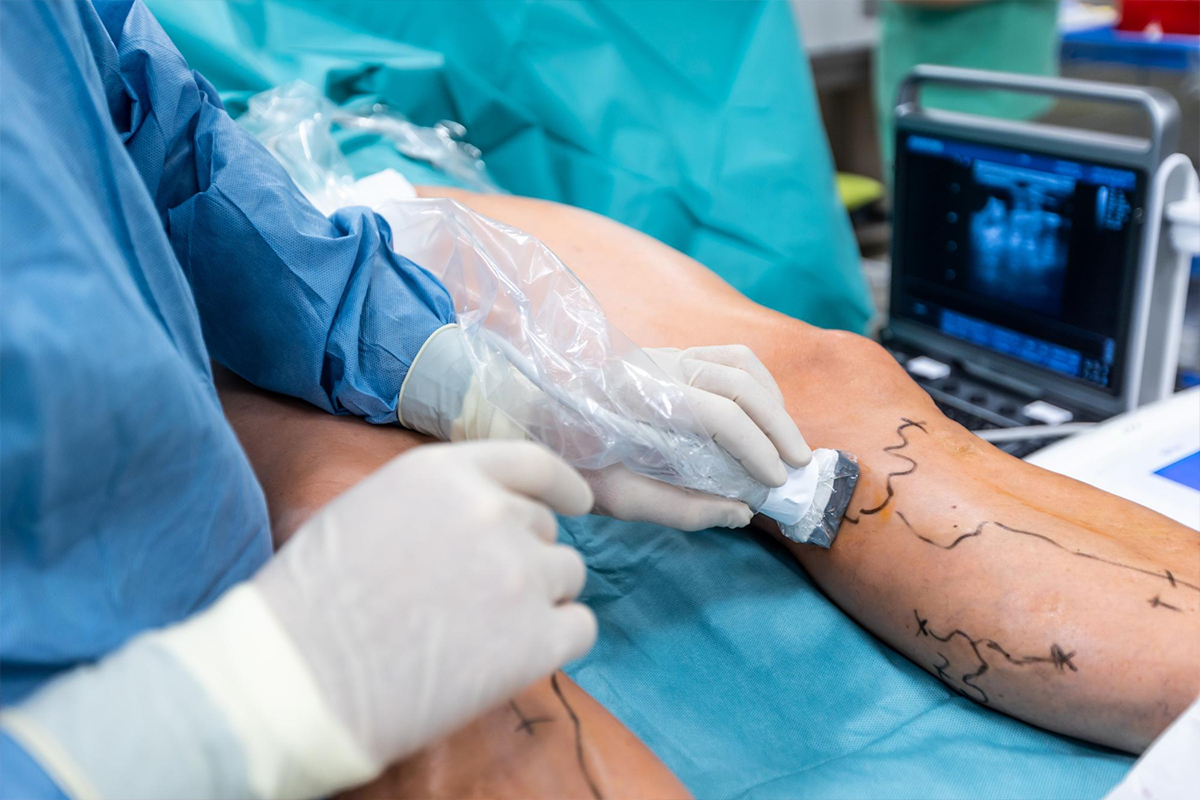
Duplex Ultrasound
-
The standard and most commonly used test. It uses sound waves to create images of blood flow and detect clots. If the vein cannot be compressed by pressure from the ultrasound probe, it likely contains a thrombus.
Venography
-
An invasive test where dye is injected into a vein and X-rays are taken to visualize clots. It is rarely used now due to the efficacy of ultrasound.
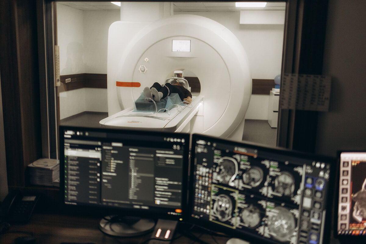
Magnetic Resonance Imaging (MRI)
-
Sometimes used to detect DVT in areas difficult to assess with ultrasound, like the pelvis or abdomen.
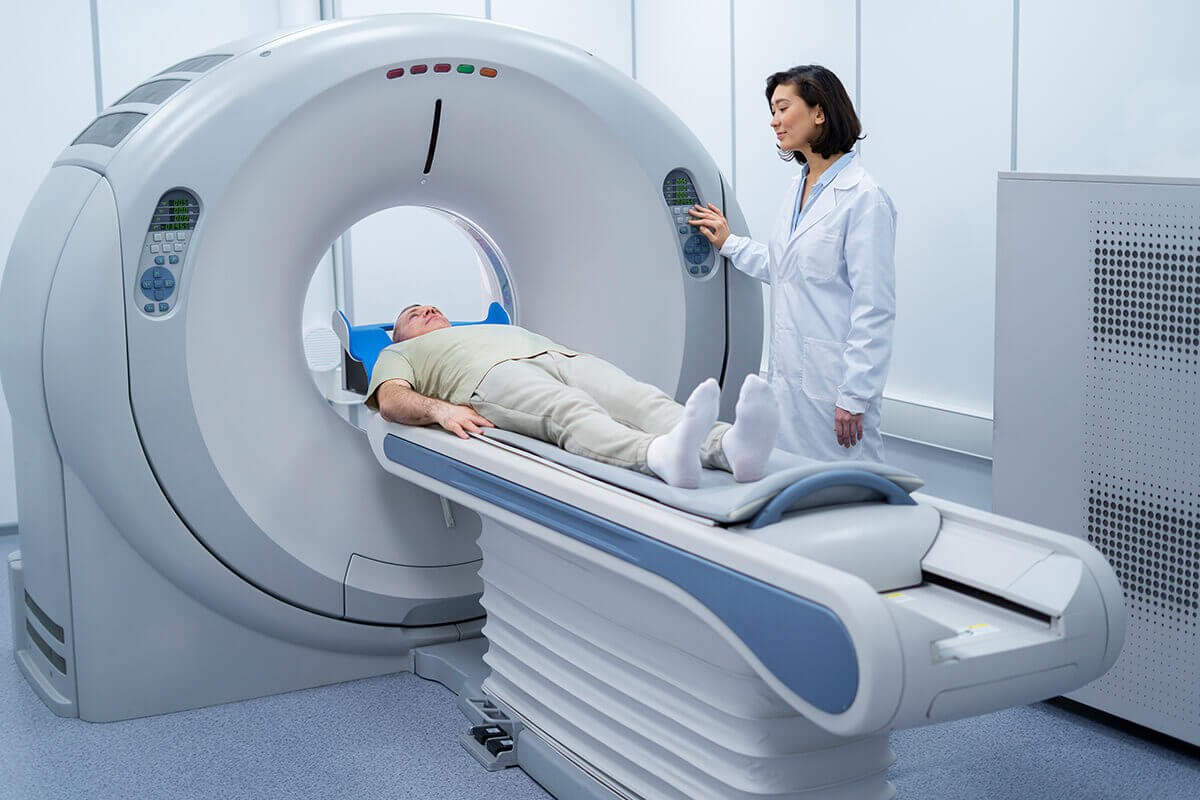
Computed Tomography (CT) Venography
-
Used in specific cases to visualize deep veins and clots.