Uterine Fibroids

Uterine Fibroids


Symptoms that get worse over time: Abdominal distension, pelvic pain, pressure symptoms (on the bladder and bowel), and heavy menstrual bleeding can all get worse and interfere with everyday activities and emotional health.
Ongoing severe bleeding can lead to chronic anemia, which can cause weakness, exhaustion, dyspnea, and heightened vulnerability to infections.
Fibroids can impede the uterus, raise the risk of preterm labor, cause miscarriages, and result in complications like fetal growth restriction and placental abruption.
Large fibroids may put pressure on the rectum (resulting in constipation), the bladder (causing frequent, urgent, or retention urination), or the ureters (causing hydronephrosis or kidney damage).
Ongoing pain and discomfort can make it difficult to go about daily tasks.
Necrosis, degeneration, and abrupt, intense pain that may necessitate emergency care can occur when fibroids outgrow their blood supply.
Hematological alterations and, in rare cases, malignant transformation may increase the risk of venous thromboembolism.

diagnosis
Lorem Ipsum is simply dummy text of the printing and typesetting industry. Lorem Ipsum has been the industry's standard dummy text. Lorem Ipsum is simply dummy text of the printing and typesetting industry

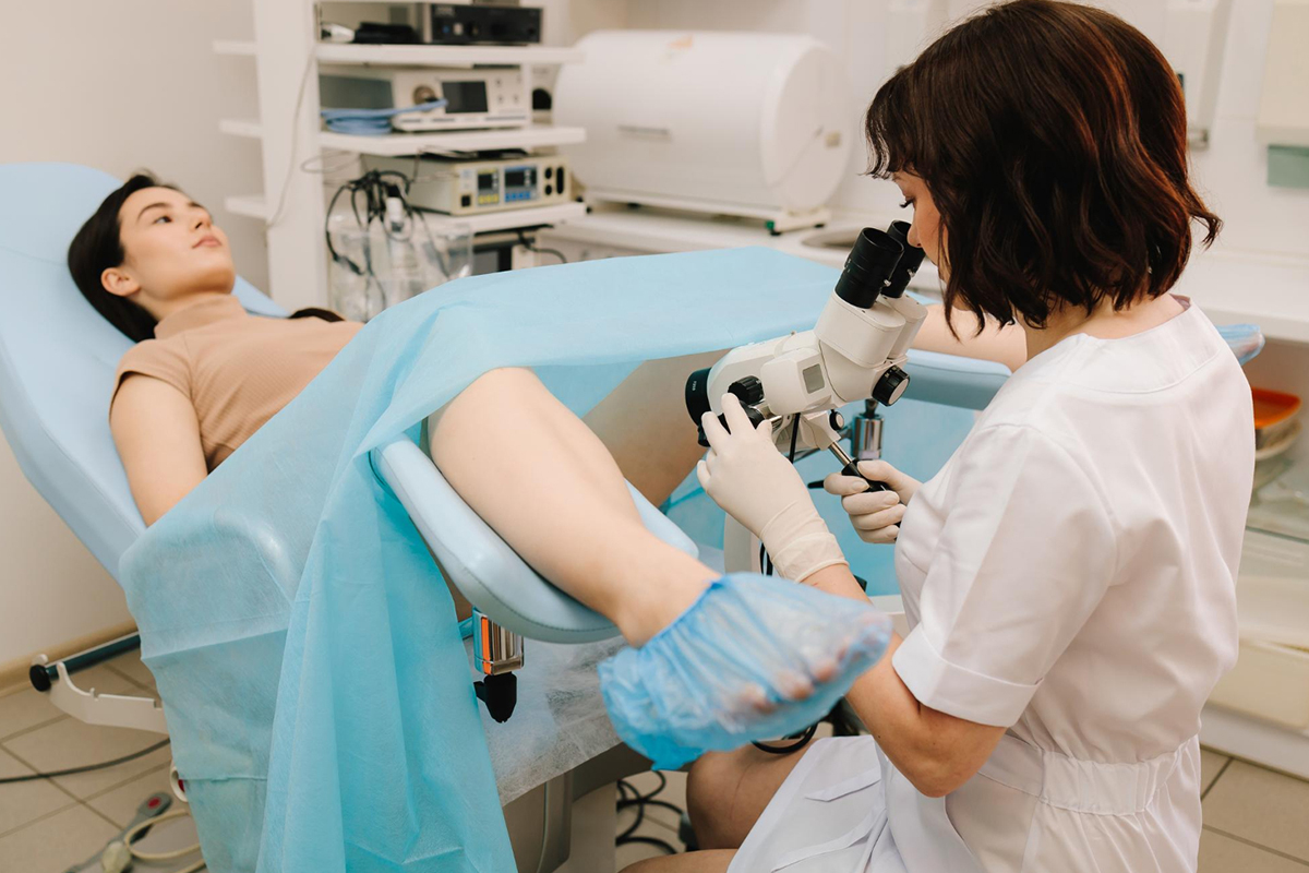
Pelvic examination
-
 During a routine pelvic exam, a medical professional may feel for an enlarged or irregularly shaped uterus, which may raise suspicions of fibroids.
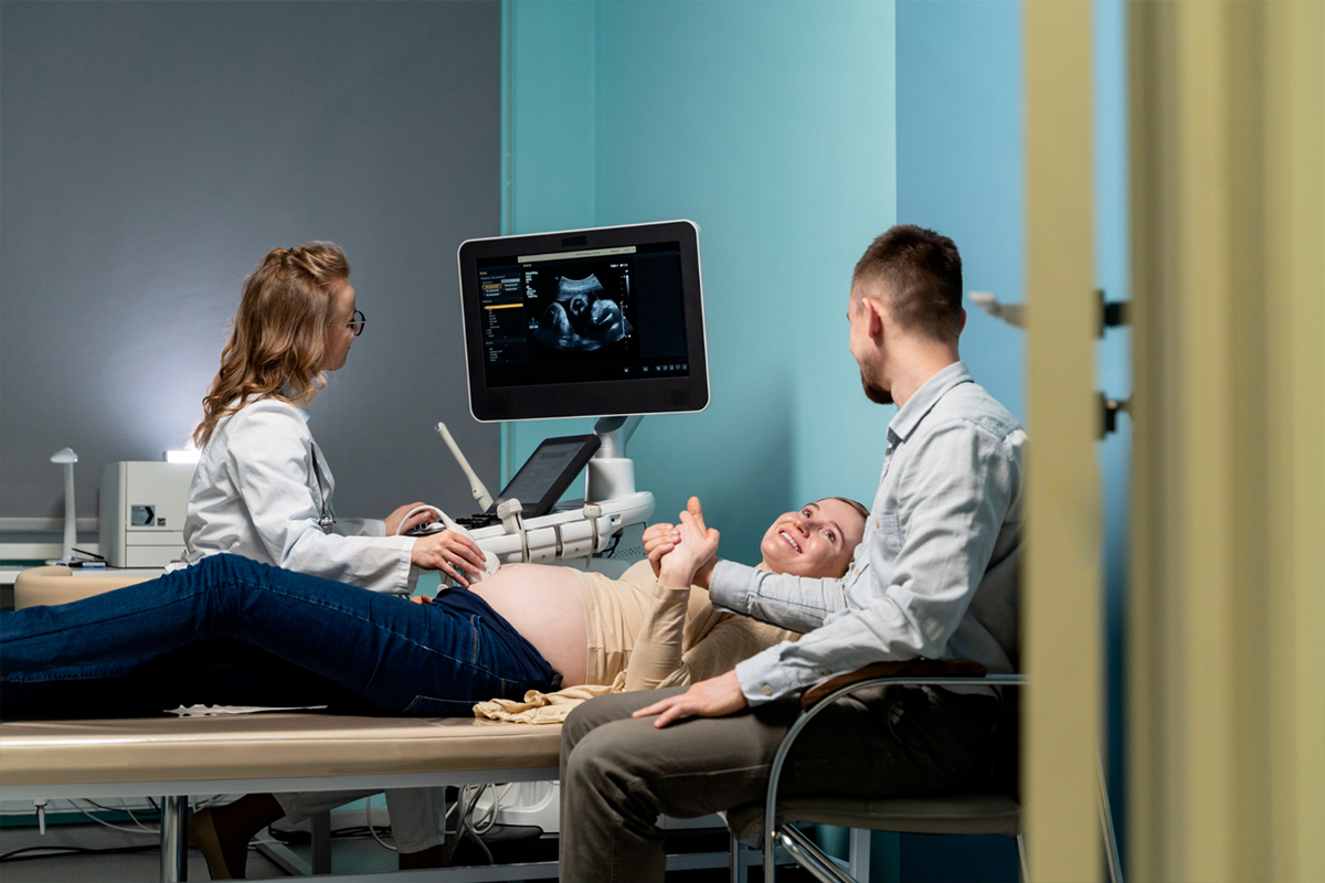
Transvaginal and transabdominal ultrasound
-
This first-line imaging test maps and measures fibroids in the uterus by using sound waves. It is widely accessible, non-invasive, and very good at finding fibroids.
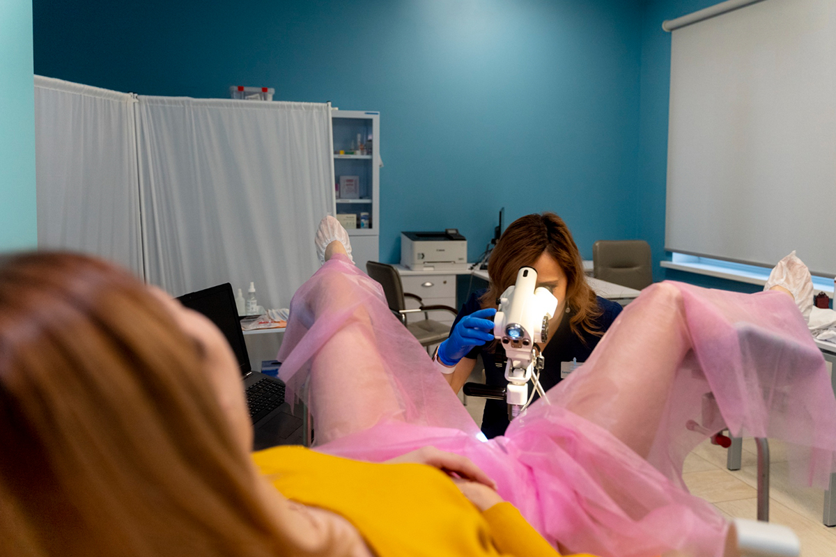
Hysteroscopy
-
To directly view the uterine cavity and submucosal fibroids, a thin instrument called a hysteroscope which combines a light and a camera, is passed through the vagina and cervix into the uterus. This process is especially helpful for assessing infertility and abnormal uterine bleeding symptoms.
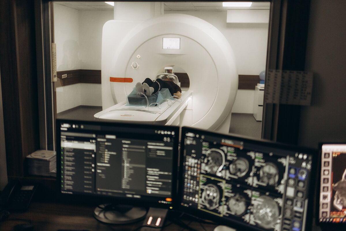
MRI
-
For large or numerous fibroids, when precise mapping is necessary for treatment planning, or when ultrasound results are unclear, magnetic resonance imaging, or MRI, is utilized. Fibroids can be distinguished from other growth types using MRI.
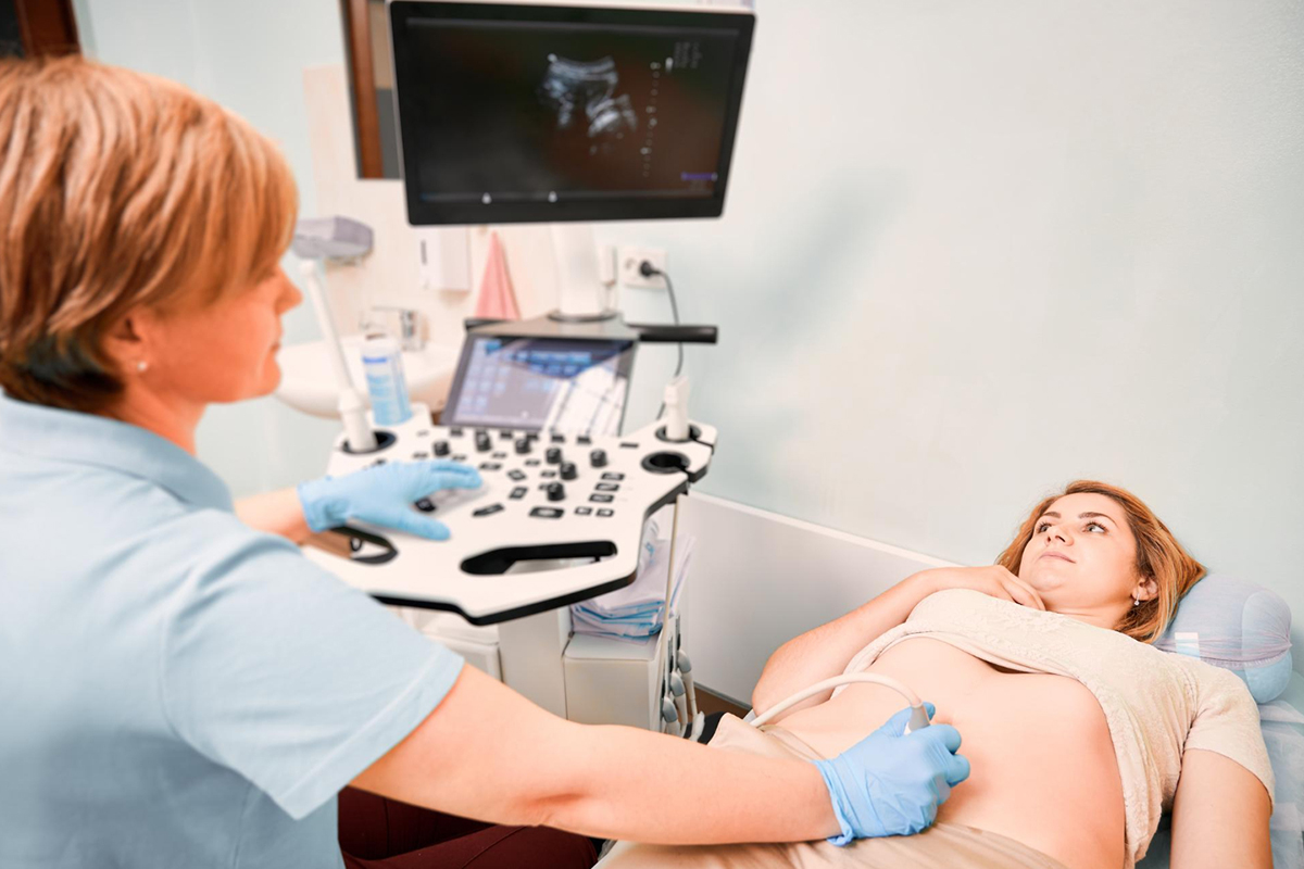
Hysterosonography
-
Saline infusion sonography, also known as hysterosonography, is a type of specialized ultrasound that uses saline to improve uterine cavity visualization and identify submucosal fibroids.
Hysterosalpingography
-
This procedure evaluates the uterus and fallopian tubes using X-rays and a dye. It is useful for identifying fibroids that protrude into these areas and for infertility workups.
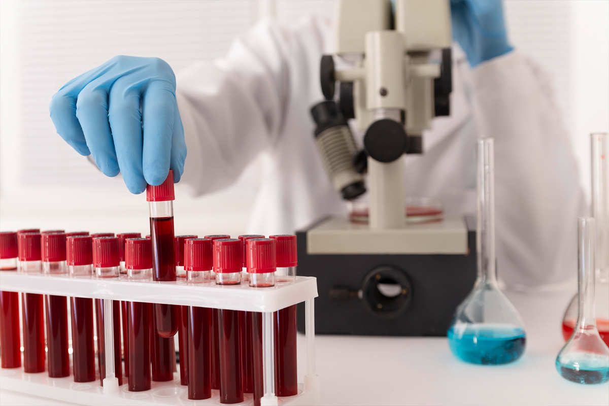
Blood tests
-
Used to check for anemia brought on by fibroids-induced heavy menstrual bleeding.