Breast Abnormalities

Breast Abnormalities


Benign lumps like fibroadenomas may remain stable or slowly grow but rarely turn cancerous.
Suspicious lumps left without biopsy or treatment pose a risk of delayed cancer diagnosis.
Cysts can cause discomfort or infection if untreated.
Larger benign lesions that grow may become painful or cosmetically concerning.
Untreated suspicious lesions can progress to malignancy, emphasizing the importance of timely intervention.

diagnosis
Lorem Ipsum is simply dummy text of the printing and typesetting industry. Lorem Ipsum has been the industry's standard dummy text. Lorem Ipsum is simply dummy text of the printing and typesetting industry

Clinical Breast Exam
-
 The first physical examination to find the lump, evaluating its size, shape, mobility, and tenderness.
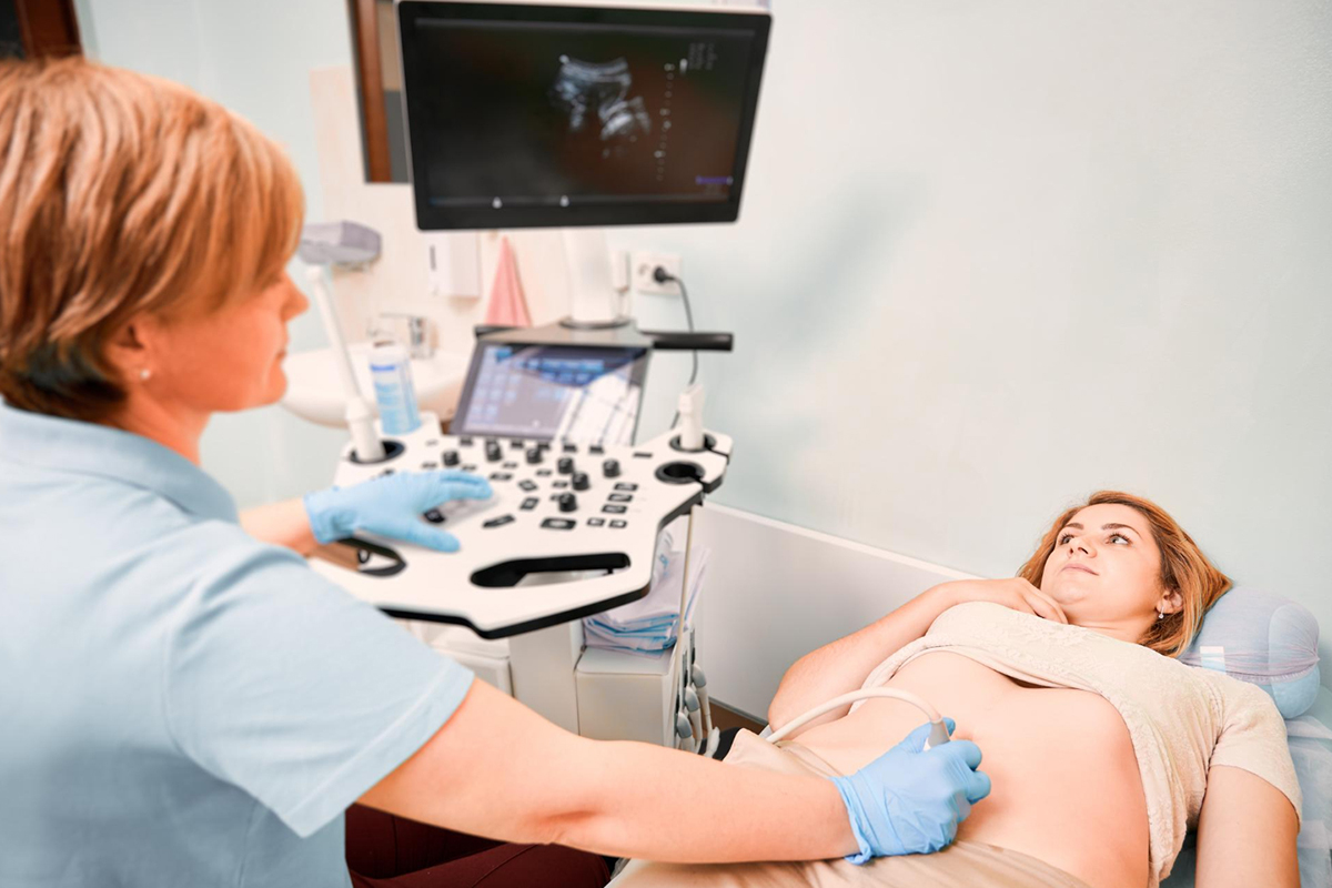
Breast Ultrasound
-
Often performed on women under 30, this non-radiative method clearly displays size and shape while differentiating between solid fibroadenomas and fluid-filled cysts.
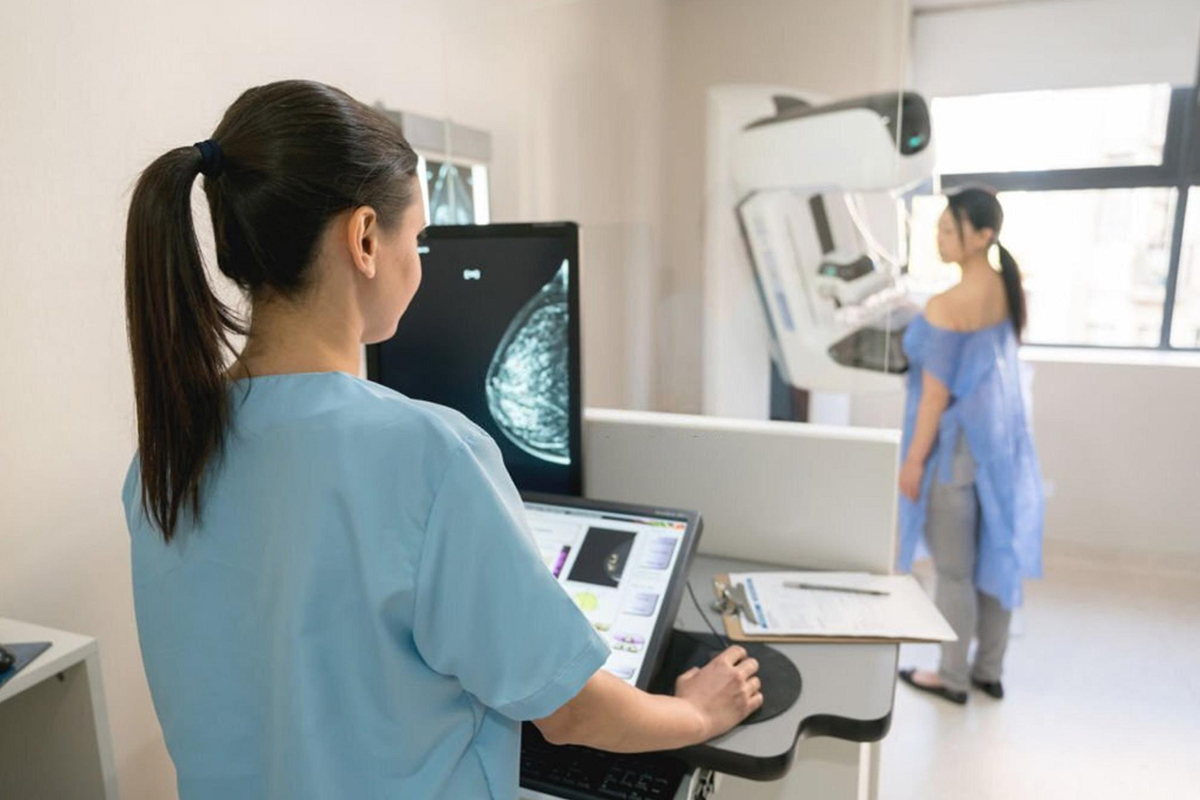
Mammography
-
Although they are less useful in younger women with dense breast tissue, mammograms, which are usually performed on women over 35, provide X-ray images to evaluate the characteristics of the lump.
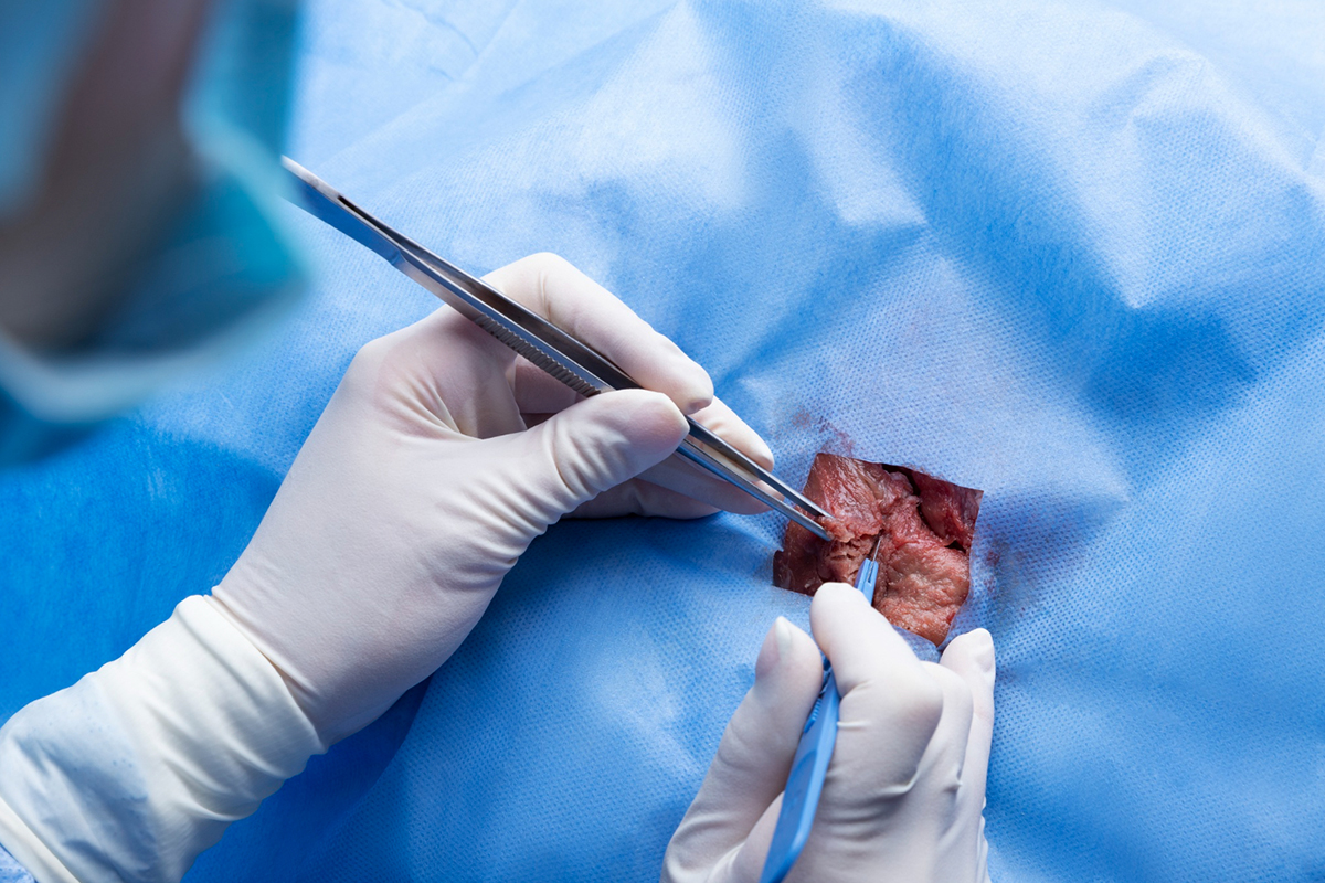
Biopsy
-
In cases where the diagnosis is unclear, tissue samples are taken for pathological analysis using a core needle biopsy, which is carried out under ultrasound guidance to confirm fibroadenoma and rule out cancer.

Fine Needle Aspiration
-
By removing cells for analysis, fine needle aspiration (FNA), a less popular technique, can assist in differentiating benign from malignant lesions.
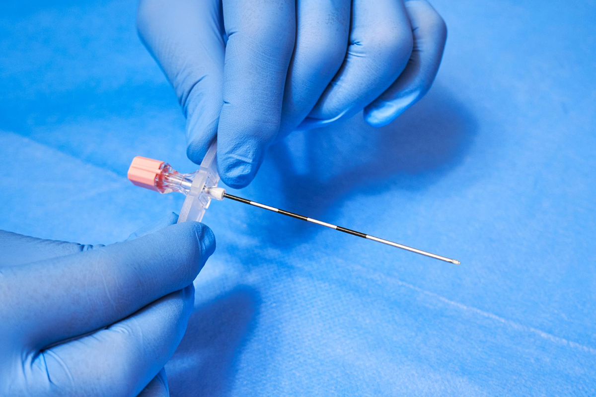
Core Needle Biopsy
-
Under ultrasound or stereotactic guidance, a needle collects tissue samples from the lesion for pathological evaluation.