Brain Aneurysm

Brain Aneurysm
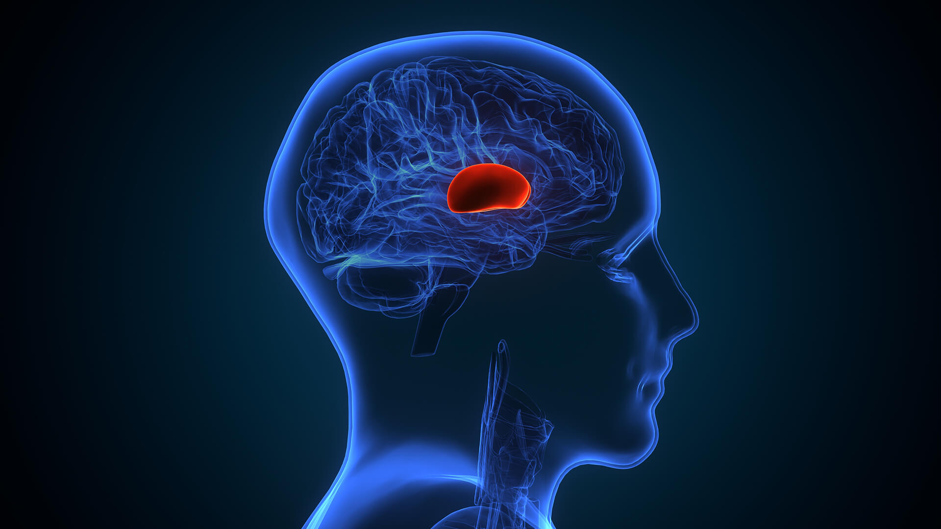

The aneurysm may burst, causing bleeding in or around the brain
Bleeding puts pressure on brain tissue, causing swelling, stroke, and irreversible neurological damage.
Survivors of rupture may suffer long-term impairments like memory loss, physical disability, seizures, speech difficulties, personality changes, and emotional disorders.
Even unruptured aneurysms can cause significant mental health issues such as anxiety and depression due to fear of rupture.
Approximately 40-50% of people with a ruptured brain aneurysm die within the first few weeks without treatment.
Untreated large or complex aneurysms can significantly shorten life expectancy.

Diagnosis
Lorem Ipsum is simply dummy text of the printing and typesetting industry. Lorem Ipsum has been the industry's standard dummy text. Lorem Ipsum is simply dummy text of the printing and typesetting industry
Cerebral Angiography (DSA – Digital Subtraction Angiography)
-
 In order to obtain detailed X-ray images of blood vessels and aneurysms, a catheter is inserted into the brain arteries and contrast dye is injected.
 -
This invasive procedure, known as cerebral angiography, is regarded as the gold standard.
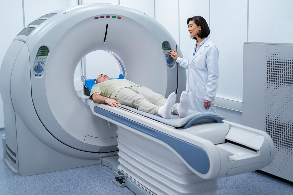
CT Scan (Computed Tomography)
-
Usually the first test performed, particularly if there is a suspicion of brain rupture and bleeding.
-
It can identify stroke or bleeding and provides two-dimensional images of the brain.
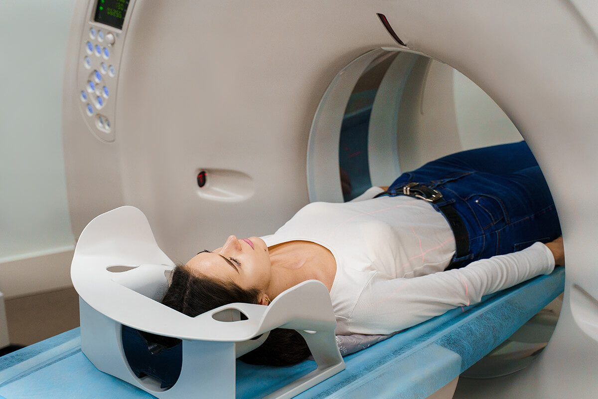
CT Angiography (CTA)
-
In order to identify and measure aneurysms, a specialized CT scan using contrast dye called CT Angiography (CTA) produces finely detailed three-dimensional images of the brain's blood vessels
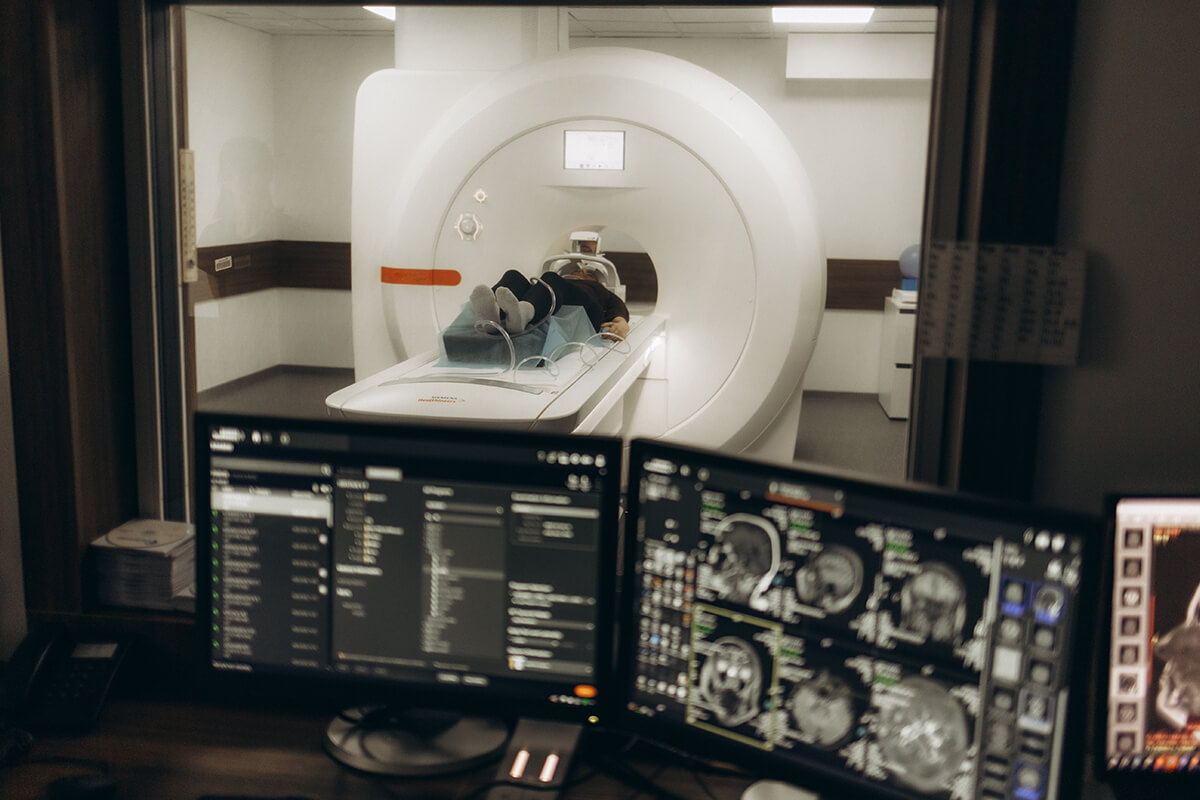
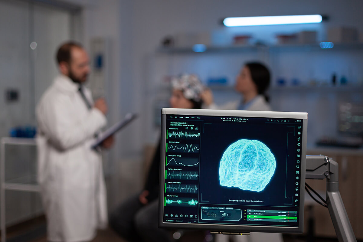
MRI / MRA (Magnetic Resonance Imaging / Angiography)
-
Creates fine-grained pictures of the brain and blood vessels using radio waves and magnetic fields.
-
A non-radiative MRI technique that focuses on the arteries in the brain to detect aneurysms and their features.

Lumbar Puncture (Spinal Tap)
-
Cerebrospinal fluid is tested for blood to diagnose subarachnoid hemorrhage caused by aneurysm rupture if a CT scan is negative but bleeding is clinically suspected.