Arteriovenous Fistulas

Arteriovenous Fistulas


Large AV fistulas cause increased blood flow that forces the heart to pump harder, potentially weakening the heart muscle and leading to heart failure.
AV fistulas, especially in the legs, can cause blood clots that may lead to deep vein thrombosis (DVT) or pulmonary embolism, which can be life-threatening.
Lack of adequate blood flow to muscles can cause pain and cramping during walking or exercise.
In cases of pulmonary AV fistulas, small clots can bypass filtration and enter the brain, causing a stroke.
AV fistulas may lead to internal bleeding, including gastrointestinal bleeding or hemorrhage in the brain.
Persistent swelling and skin discoloration near the fistula site.

diagnosis
Lorem Ipsum is simply dummy text of the printing and typesetting industry. Lorem Ipsum has been the industry's standard dummy text. Lorem Ipsum is simply dummy text of the printing and typesetting industry

Physical Examination
-
 A healthcare provider listens with a stethoscope for a continuous humming or whooshing sound (bruit) near the fistula and feels for a vibration (thrill) over the artery and vein connection.
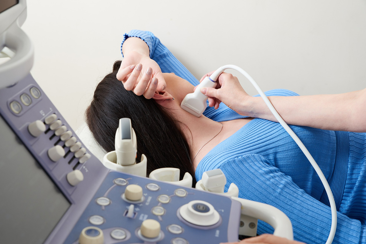
Duplex Ultrasound
-
The most common, non-invasive imaging test that uses sound waves to visualize blood flow and detect abnormal connections between an artery and a vein.
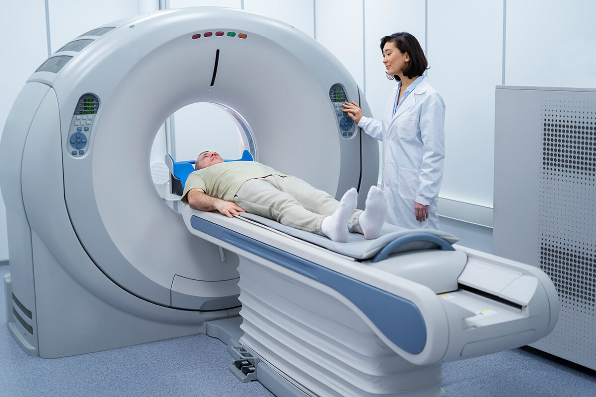
Computed Tomography (CT) Angiogram
-
Uses X-ray images and contrast dye to create detailed pictures of blood vessels and identify fistulas, especially when located deeper.
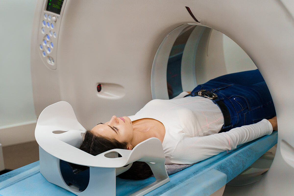
Magnetic Resonance Angiography (MRA)
-
Uses magnetic fields and radio waves with contrast dye to image blood vessels and diagnose fistulas without radiation exposure.
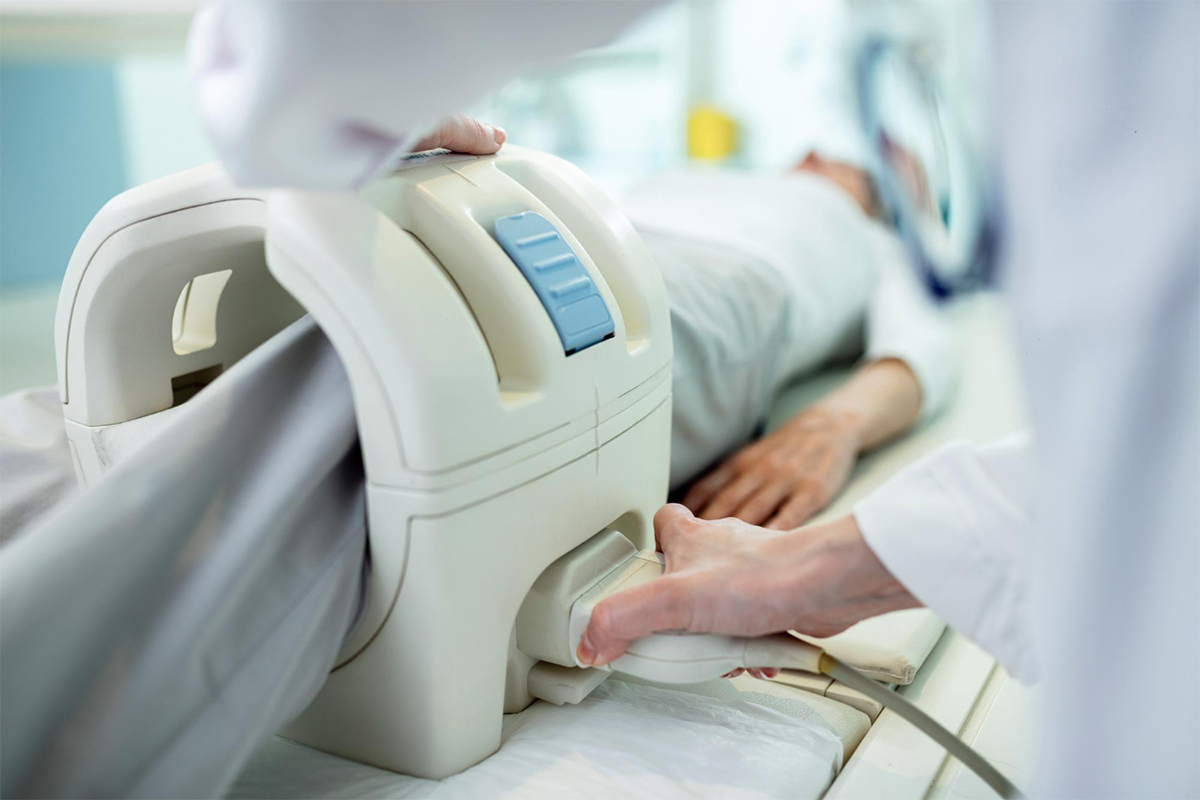
Conventional Angiography
-
An invasive procedure where contrast dye is injected directly into the blood vessels to provide precise imaging; often considered the gold standard for detailed mapping before treatment.

Additional Tests
-
Blood flow measurements and oxygen content assessments may be used for assessing fistula function and hemodynamics.