Spinal Vascular Malformation
WHAT IS
SPINAL VASCULAR MALFORMATION?
Spinal vascular malformations are abnormal clusters of blood vessels within or near the spinal cord, which can disrupt blood flow, leading to pain, weakness, numbness, or even paralysis if left untreated.
causes
Congenital abnormalities in blood vessel development
Genetic/hereditary factors
Associated with neural tube defects
Trauma or injury to spinal blood vessels
Possibly acquired or idiopathic in some adult cases
symptoms
Trouble walking or climbing stairs
Numbness, tingling, or sudden pain in the legs
Weakness on one or both sides of the body
Sudden, serious back pain
Loss of sensation or numbness in the legs
Difficulty urinating or bowel movement problems
Imbalance and coordination difficulties
Sexual dysfunction
Headache and neck stiffness
Progressive spinal cord dysfunction leading to paralysis
if untreated?

Progressive disability worsening over time

Permanent spinal cord damage

Weakness or paralysis

Pain, tingling, and numbness

Spinal cord infarction (tissue death due to lack of oxygen)

Hemorrhage or bleeding in the spinal cord

Bulging blood vessels (aneurysms)

Venous hypertension causing fluid buildup and tissue damage

Loss of bladder and bowel control

Potentially severe neurological impairments or death
diagnosis
Lorem Ipsum is simply dummy text of the printing and typesetting industry. Lorem Ipsum has been the industry's standard dummy text. Lorem Ipsum is simply dummy text of the printing and typesetting industry
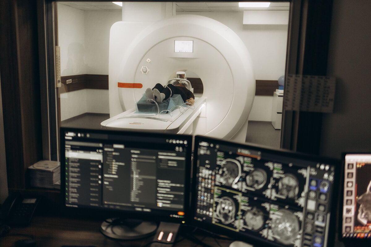
Spinal MRI
-
The first and most popular test for identifying abnormal blood vessels and spinal cord abnormalities is spinal MRI.
Spinal angiography
-
The gold standard for confirming a diagnosis is spinal angiography, also known as digital subtraction angiography, which provides the exact location and specifics of the malformation.
Spinal MRA or CTA
-
Less detailed than angiography, but useful as screening tests to see blood vessels.
Physical examination and symptom evaluation
-
Assess symptoms and perform a physical examination. Use neurological deficits to guide imaging studies.
Interventional Radiology
treatment
Embolization using Onyx or glue agents
In order to block blood flow through the abnormal vessels, a microcatheter is inserted into the AVM’s feeding arteries and liquid agents, such as Onyx or special medical glue, are injected. This minimally invasive procedure is known as embolization. Embolization is frequently used as part of a combination treatment plan to help shrink or close the AVM, increasing the safety or efficacy of radiosurgery or surgery.
Stereotactic Radiosurgery
With stereotactic radiosurgery, the AVM is precisely targeted without requiring open surgery using intensely concentrated radiation beams, which are frequently delivered by tools like a Gamma Knife or CyberKnife. When surgery is not an option for small or deep AVMs, radiosurgery is frequently used to damage the vessel walls of the AVM, causing them to gradually close off. Gradual obliteration usually happens months to years after treatment.
Stent-assisted coiling
In cases of complex or wide-necked spinal aneurysms and fistulas, a stent is inserted inside the vessel to support coil placement and stop coil migration.
Balloon-assisted coiling
A tiny balloon is temporarily inflated across the opening of the vessel during coil placement to hold coils in place; used in challenging anatomies for improved coil stability during embolization.
