May-thurner Syndrome

May-thurner Syndrome
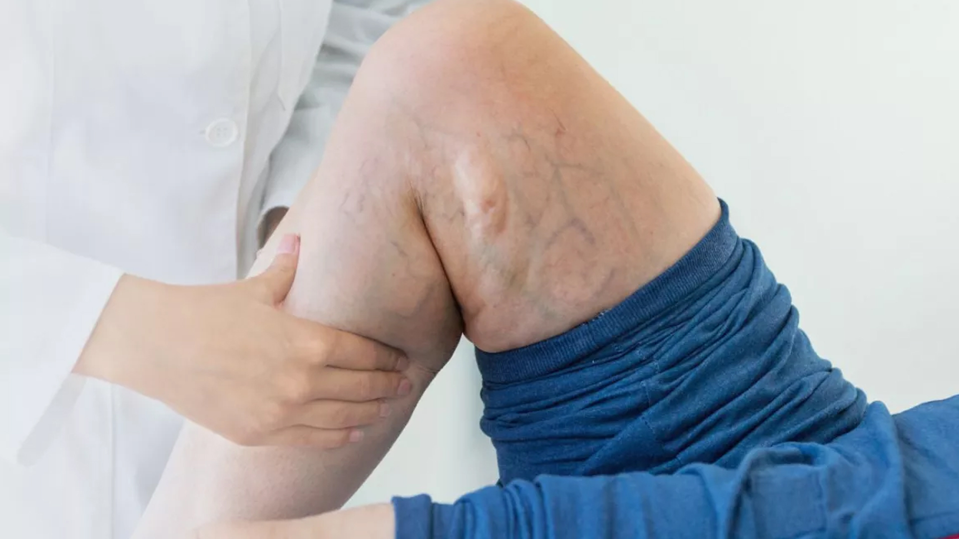

Deep vein thrombosis (DVT) developed in the left leg as a result of impaired blood flow and persistent venous compression.
A pulmonary embolism (PE) is a potentially fatal blockage that happens when a blood clot from the leg breaks loose and moves to the lungs.
Blood buildup in the veins, known as chronic venous insufficiency, results in chronic leg swelling, pain, skin changes, and non-healing venous ulcers.
Persistent skin discoloration, swelling, and leg pain that severely lowers quality of life.
Higher chance of blood clots happening again and potential consequences like heart attacks or strokes if the clots spread to other organs.

diagnosis
Lorem Ipsum is simply dummy text of the printing and typesetting industry. Lorem Ipsum has been the industry's standard dummy text. Lorem Ipsum is simply dummy text of the printing and typesetting industry
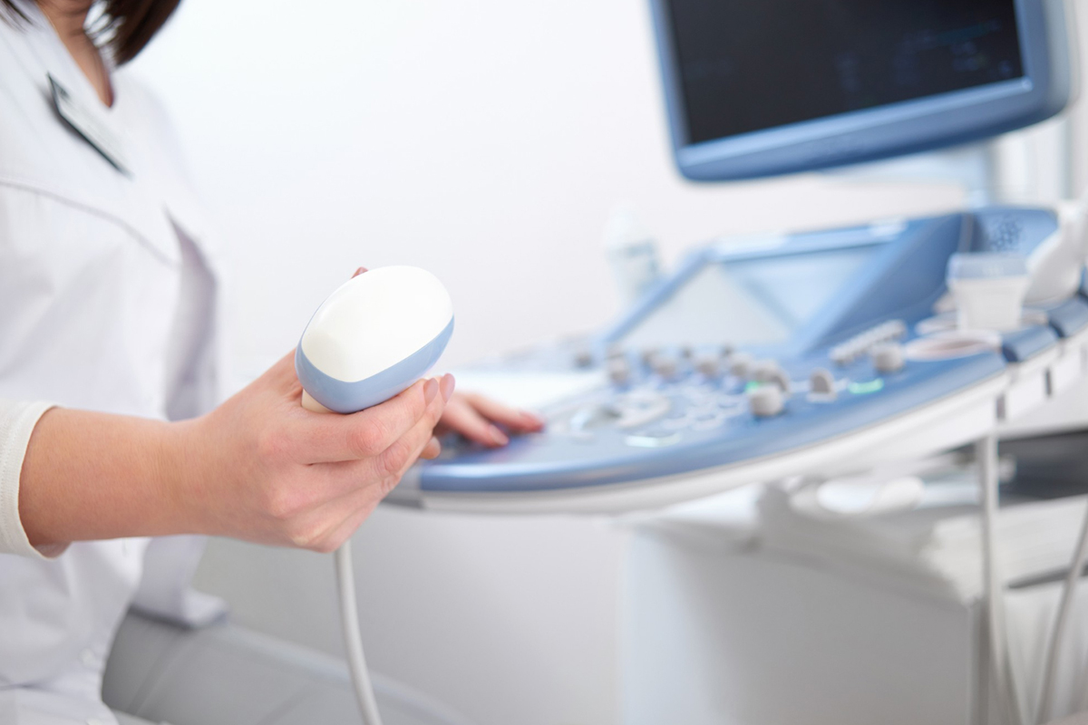
Doppler Ultrasonography
-
 This is frequently the first non-invasive test to measure venous reflux, identify thrombosis, and gauge blood flow in the iliac veins. However, because of its deep pelvic location and surrounding structures, it might have trouble seeing the iliac vein.
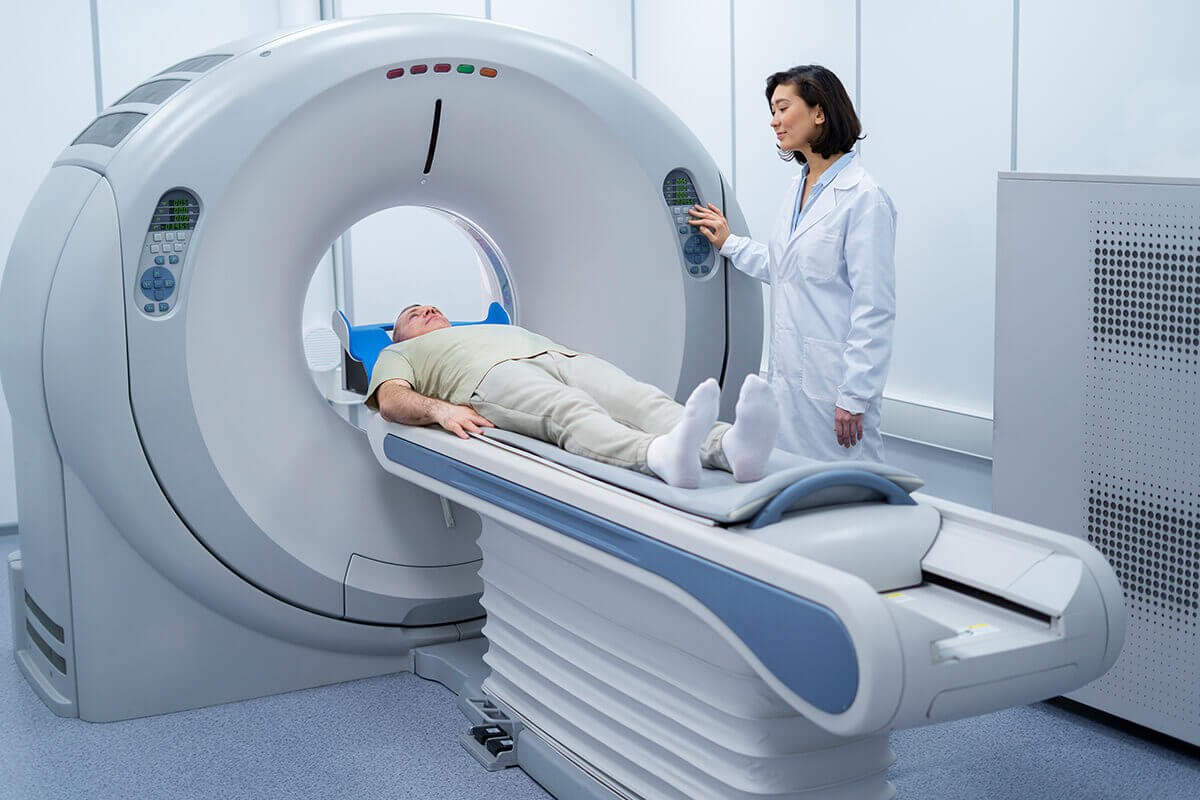
Computed Tomography Venography (CTV)
-
Offers fine-grained cross-sectional pictures that demonstrate the presence of collaterals, thrombus, and other pelvic anomalies as well as the compression of the left iliac vein by the right iliac artery. It can diagnose MTS with high sensitivity and specificity.
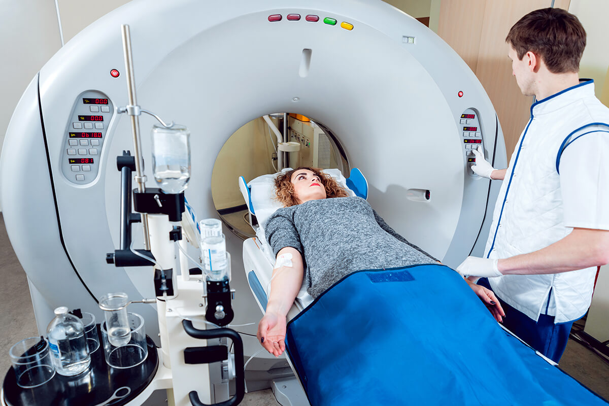
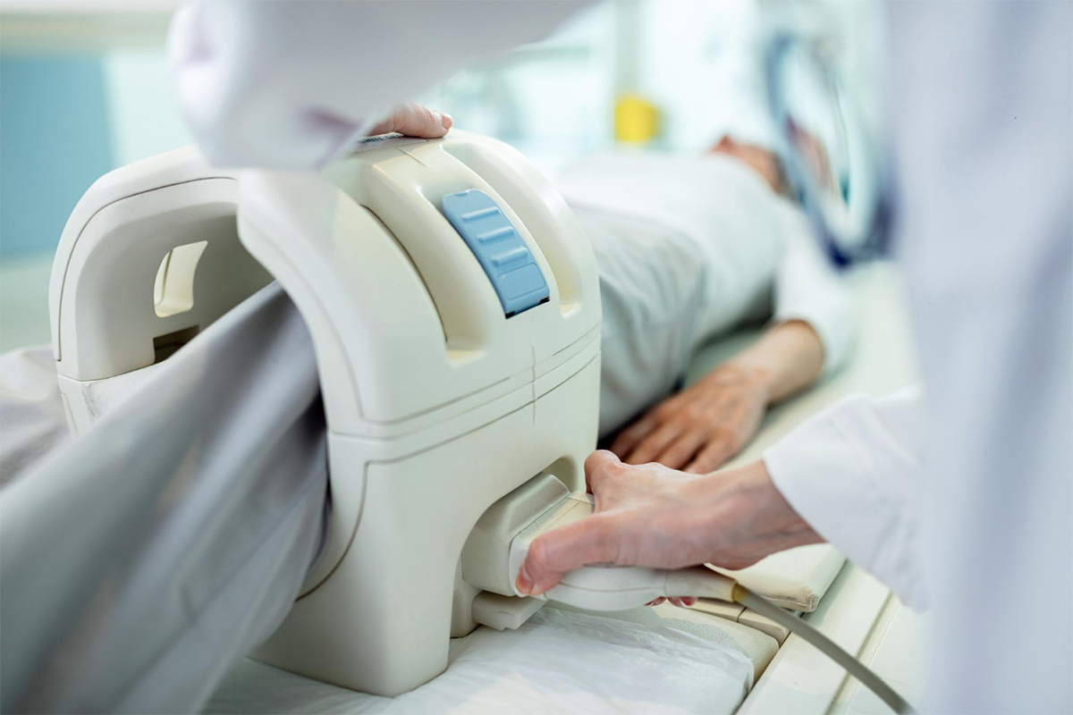
Magnetic Resonance Venography (MRV)
-
A radiation-free substitute for CTV that is particularly helpful during pregnancy. Venous compression, collateral veins, and the distinction between thrombus and flow artifacts can all be seen with MRV.
Conventional Catheter Venography
-
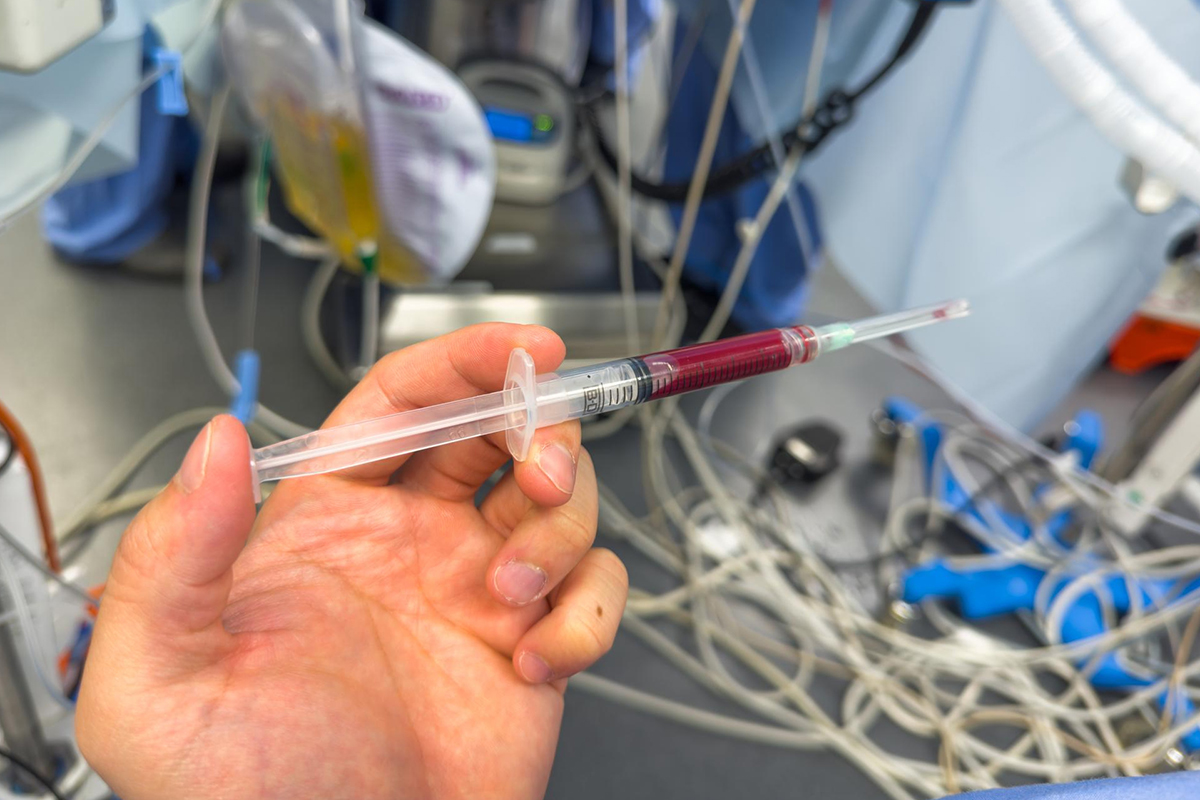
The gold standard for diagnosis is conventional catheter venography, also known as digital subtraction venography, or DSV. In order to view the iliac veins and detect stenosis or occlusion, a catheter-based contrast injection is used. Additionally, it is capable of measuring pressure gradients throughout the constricted area.
Intravascular Ultrasound (IVUS)
-
Often utilized in venography or intervention, IVUS helps with precise stent placement, detects intraluminal spurs and webs, and provides real-time, high-resolution images of the vein lumen and wall.

Physical examination and clinical history
-
If there are no other explanations for the swelling and pain in the left leg, it may raise suspicion and lead to imaging tests.