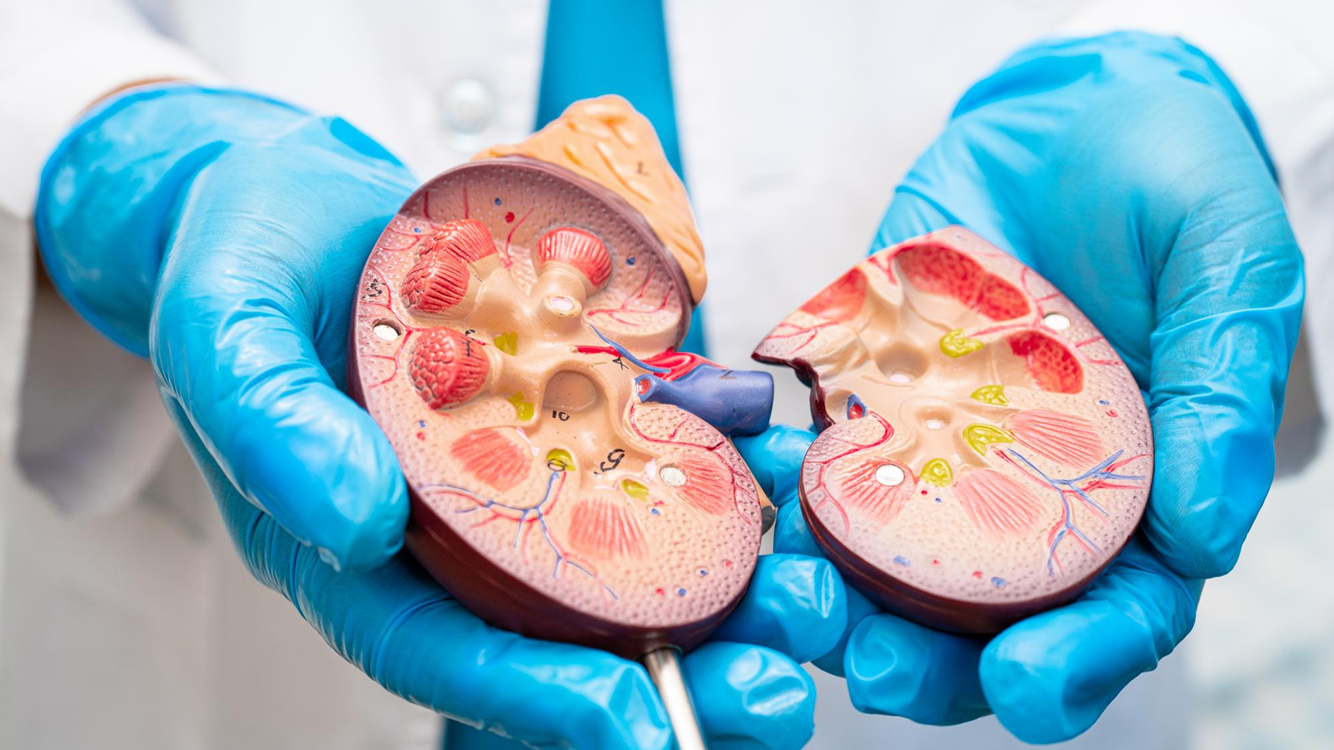
Renal Tumors

Renal Tumors

Untreated kidney cancer can grow and invade nearby tissues and organs.
It can spread (metastasize) to distant organs like lungs, bones, and brain.
Symptoms worsen, including pain, blood in urine, weight loss, fatigue, and swelling.
Advanced disease may cause organ failure and severe complications.
Median survival for untreated advanced kidney cancer is often less than a year.
Early treatment significantly improves survival and reduces symptoms.

diagnosis
Lorem Ipsum is simply dummy text of the printing and typesetting industry. Lorem Ipsum has been the industry's standard dummy text. Lorem Ipsum is simply dummy text of the printing and typesetting industry
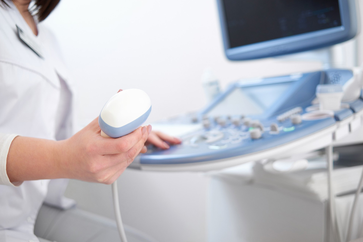
Imaging tests such as ultrasound
-
 Imaging tests such as ultrasound, CT scans, and MRI are used to visualize the kidneys and detect masses.

Ultrasound
-
Ultrasound helps differentiate solid tumors from cysts.
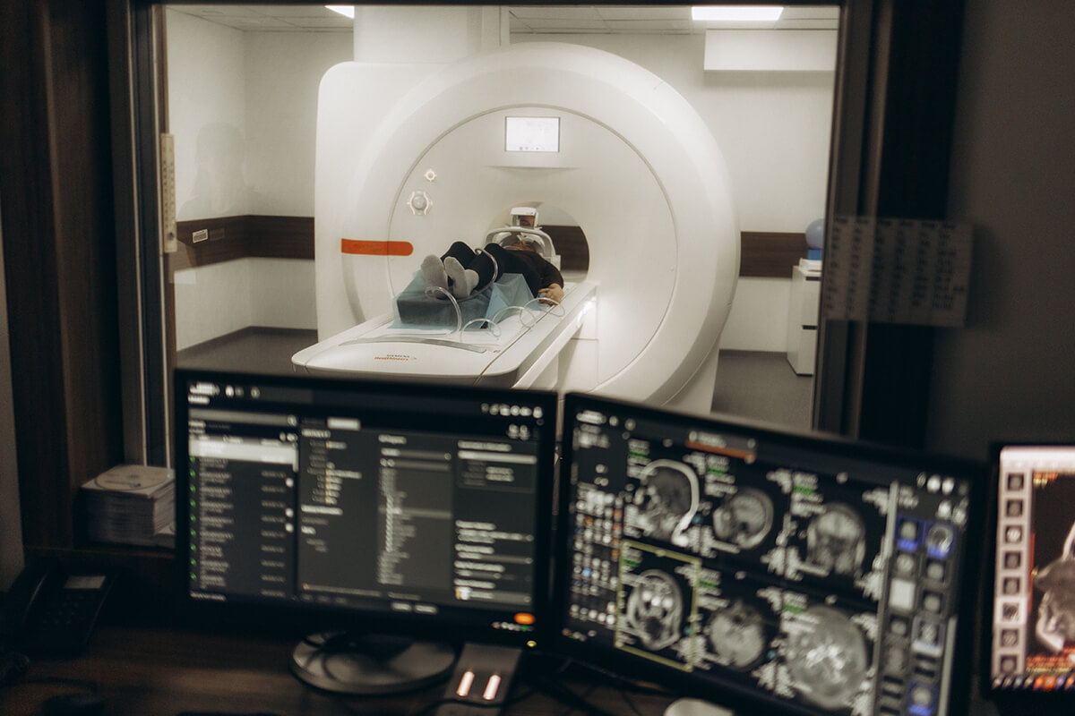
CT and MRI
-
CT and MRI provide detailed cross-sectional images showing tumor size, location, and spread to lymph nodes or other organs.

Blood and urine tests
-
Blood and urine tests assess kidney function and detect signs like anemia or blood in urine but do not confirm cancer.

Percutaneous needle biopsy
-
Percutaneous needle biopsy is done when imaging is inconclusive, taking a small tissue sample for microscopic examination.

X-rays or bone scans
-
Additional tests like chest X-rays or bone scans check for metastasis.